


ICCK Journal of Image Analysis and Processing
ISSN: 3068-6679 (Online)
Email: [email protected]

Capturing images in everyday life is that it helps remember memories. Taking images allows you to capture special moments, everyday occurrences, and the evolution of people and places over time [1]. An ideal image should perfectly capture every detail of a scene, providing all the necessary information. However, since cameras have limitations, this isn't always possible [2]. One of the most common photographic challenges is to capture an image with varying focal depths, where other parts of the scene have become focused, as well as parts that have been blurred or unclear. This is a result of the fact that the lens of a camera cannot focus at a different depth. Image fusion techniques are employed to overcome this problem [3].
Image fusion combines elements from multiple photos into a single picture that captures every detail of the scene clearly. This technique is particularly useful when a camera's low depth of field avoids it from capturing everything in focus at once. For example, when taking a photo, the camera may focus well on either a nearby or a distant object, but not both. Image fusion addresses this problem by combining images taken at different distances [4]. Image fusion is used in many different kinds of applications to improve image clarity and information content. In medical imaging, it combines data from various scans such as MRI and CT to improve diagnostic accuracy. Remote sensing uses images from multiple spectral bands to improve monitoring of the environment. It is also used in monitoring to combine feeds from multiple cameras for complete monitoring, and in automotive technology to help advanced driver-assistance systems by consolidating sensor data to improve safety features [5].
Multifocus image fusion is an approach for combining a set of images that are differently focused, to form a single fused composite image and clear representation such that all useful information in every image is well-focused ensuring the efficiency on further analysis or visualization not limited by any loss occurred from defocusing [6]. Multifocus image fusion is majorly used in medical imaging for sharp diagnostic visuals, and improved surveillance so clearer image and photography to achieve high depth of field [7].
In this article, we explore the application of image fusion techniques consisting of Marr–Hildreth edge detection, Discrete Cosine Transform (DCT), Stationary Wavelet Transform (SWT), and Discrete Wavelet Transform (DWT) to perform image fusion, especially multi-focus one. The Marr–Hildreth technique enhances image features by applying a Gaussian blur followed by the Laplacian operator, which helps in detecting and preserving edges. The Discrete Cosine Transform (DCT) is used for separating the image into parts of varying importance, increasing efficiency by focusing on the more important components [8]. Furthermore, the Stationary Wavelet Transform (SWT) helps in maintaining shift-invariance, ensuring better structural preservation in images, while the Discrete Wavelet Transform (DWT) provides efficient multi-resolution decomposition, allowing images to be analyzed at different levels of detail, which is important for effectively combining data from multiple sources [9].
The first step involves collecting two multi-focused images and applying Marr–Hildreth edge detection as a preprocessing technique. This method enhances image sharpness by detecting and preserving edges, which contributes to better fusion results. After preprocessing, the images are finally combined using image fusion techniques such as DCT, SWT, and DWT to get a single clear, enhanced composite image. The objective is to produce a final image that enhances significant information from the original elements, improving the quality of the total image.
The dataset for this experiment contains four groups of grayscale photos: "Balloons," "Leaves," "Clock," and "Clocks and Leaves." These collections are based on the "Lytro multi-focus datasets" [10]. They have been selected specifically to improve the evaluation and testing of multi-focus images using modern techniques. This study made the following contributions:
The images were pre-processed using the Marr–Hildreth edge detection technique to enhance image clarity and sharpness. This method involves applying a Gaussian blur to the image to reduce noise, followed by computing the Laplacian to detect areas with rapid intensity changes, effectively highlighting edges and fine details.
The proposed method used advanced image fusion methods DCT, SWT and DWT that combines multiple images captured at different focal depths to improve image clarity and detail. The experiments also discuss the fusion results using different datasets.
The experiments were designed to check the efficiency of multi-focus image fusion on a dataset that includes four sets of grayscale images ("Clocks" "Balloons," "Leaves," and "Clock and Leaves"). This testing is important for calculating the effectiveness of the image fusion techniques used.
The paper is structured as follows: Section 2 covers previous research. In Section 3, we describe our research methodology. Section 4 describes the datasets, and Section 5 presents the results and discussions. Finally, Section 6 summarizes the findings and outlines future directions of the study.
Multifocus image fusion implies combining images taken at various focus levels to achieve one focused image, where details are well-defined all over. The development has taken place from simple pixel-based techniques to better synthesis algorithms, including wavelet transforms and neural networks. Applications have been noted in various imaging fields, including medical images, remote sensing, and photography, for clarity and detail to help with better analysis and interpretation. Different researchers worked in the area of multifocus image fusion such as Liu et al. [11] suggested method for a new Algorithm of Multi-Focus Color Image Fusion Based on Low-Vision Image Reconstruction and Focused Feature Extraction. They proposed an architectural gradient-based method to determine the focused feature and how it can be extracted to increase the image resolution in the multi-focused point scenarios. One of the main novelties in their approach was a deep residual network, together with a degradation model designed to further improve the level of detail in the fused images. The algorithm accurately detected areas of focus and realized a huge progress in keeping the image's detail and edge structures well preserved, compared to the existing techniques.
Li et al. [12] introduced a swift and efficient image fusion approach that strategically merges multiple images to generate a single, highly informative composite. This method hinges on a dual-scale decomposition framework, where an input image is separated into a base layer—capturing large-scale intensity variations—and a detail layer—responsible for finer structural elements. To achieve effective fusion, a novel guided filtering-based weighted average algorithm is employed, ensuring robust spatial consistency throughout the fusion process. Extensive experiments involving multispectral, multifocus, multimodal, and multiexposure image datasets underscore the superior performance of this technique, as it consistently delivers fused images of remarkable visual clarity and detail preservation across diverse application scenarios.
Zhang et al. [2] presented a comprehensive and detailed survey of multi-focus image fusion (MFIF), specifically addressing the advancements and limitations of deep learning-based methods that have emerged in this field since 2017. Recognizing the gap in the existing literature—namely, the absence of an exhaustive review focusing on deep learning for MFIF—this paper thoroughly reviews a broad array of algorithms, datasets, and evaluation metrics employed in contemporary MFIF research. As the first of its kind, this survey not only charts the progress of deep learning-driven MFIF approaches but also systematically compares them with traditional fusion techniques, highlighting significant improvements in performance as evidenced by both qualitative and quantitative metrics. Furthermore, the authors provide insightful commentary on the present state of MFIF and outline promising directions for future investigations, thereby offering a valuable roadmap for further research and applications in high-detail imaging domains.
Turgut et al. [13] used a gradient transform-based novel method to combine the focused and unfocused pixels. This aimed to counter the common problems witnessed within the performance-hindering already in use methods, such as shift variance and misregistration. It starts with generating halftone images using the Halftoning-Inverse Halftoning (H-IH) transform. The focus measurement is calculated for these gradient images through the Energy of Gradient (EOG) and Standard Deviation functions, forming a composite image. This composite was further improved with a decision fusion strategy using majority voting for the combination. The method was at the time very unique to most common robustness issues in fusion and, at the same time, captures the details which are normally unobservable by other methods. This new innovative approach has shown excellent results compared to 17 different modern and traditional techniques, both visually and objectively. In a more objective manner, the assessments were using six quantitative metrics, where with the new method doing better in five out of the six, pointing to a huge improvement over other technique.
Li et al. [6] proposed a robust and effective deep learning-based image fusion approach designed to address the challenges of integrating infrared and visible images into a single composite image with all essential features preserved. The technique initiates with a decomposition of the input images into base and detail components, enabling a structured and balanced fusion. Specifically, the base parts of the images are merged through a weighted averaging process to maintain global structure, while the detail content is refined using a deep learning network to extract multi-layer features that encapsulate rich spatial and spectral information. To further improve the fused detail quality, a combination of -norm and weighted-average strategies is utilized to generate multiple candidate fused detail contents, followed by a max selection step to finalize the fusion process. Ultimately, the fused image is reconstructed by integrating the refined base and detail components. Experimental evaluations underscore the approach's superior performance in both visual quality and quantitative metrics, positioning it as a promising tool for high-detail image fusion applications.
Zhou et al. [3] presented a comprehensive review of multi-focus image fusion (MFIF), a critical technique in overcoming the depth of field limitations inherent in optical lenses by merging multiple partially focused images into a single, fully focused composite. The authors systematically introduced a new classification system for MFIF algorithms, categorizing them into four distinct groups: transform domain methods, boundary segmentation methods, deep learning-based methods, and hybrid fusion approaches. Within this framework, they elaborated on the defining characteristics of boundary segmentation methods, highlighting their effectiveness in preserving edge information and maintaining image integrity compared to conventional methods. The paper further delineates both subjective and objective evaluation metrics, offering an in-depth discussion of eight widely recognized objective indicators used to assess the quality of fusion results. Drawing on an extensive body of literature, the authors compared and summarized a variety of representative algorithms, providing a well-rounded overview of the field. The work concludes by identifying the primary challenges faced by current MFIF research and outlining future directions for development, underscoring the method's potential for advanced applications requiring high levels of detail and visual clarity, such as medical imaging and remote sensing.
Kaur et al. [14] explored the intrinsic limitations of optical lenses, which, constrained by finite depth-of-field (DOF), struggle to produce fully focused images in a single capture. In practice, only elements within the DOF appear sharp, leaving other regions blurred. To address this challenge, researchers have proposed a suite of algorithms aimed at fusing multiple partially focused images of the same scene into a single, fully focused composite. The authors systematically reviewed these algorithms, classifying them into conventional and deep learning-based approaches, while emphasizing the unique challenges of multi-focus image fusion (MFIF). Applications of MFIF are diverse and impactful, spanning optical microscopy, micro-image fusion, digital photography, and remote sensing networks. Beyond outlining the conceptual and algorithmic underpinnings, this paper performed a comprehensive qualitative and quantitative evaluation of seven representative MFIF methods using the Real-MFF dataset. The findings highlighted both the advancements and current limitations of existing approaches, culminating in a forward-looking discussion on promising directions for future research and development in the field. Pan et al. [15] suggested an innovative methodology that is DDMF, as denoted by the Feature Difference Network for the betterment of Multi-Focus Image Fusion MFIF. The technique introduces noise to highlight the difference in the information loss between the focused and defocused regions of an image. The method applies Gaussian noise to the picture to improve the representation of features at the denoising stage while using a Denoising Diffusion Probabilistic Model diffusion process. This enables DDMF to detect and label each pixel much more accurately. Their experiments show that this new approach outperforms existing methods in terms of visual quality and objective performance measures.
You et al. [16] introduced new multi-focus image fusion technique by applying the local standard deviations of Laplacian images from the input images that are further enhanced with a guided filter. The key innovation is that locally computed standard deviation indicates the sharpness of the images; indeed, if the pixel is sharper, then the local variance is higher. This technique includes the application of Laplacian filters to source images, followed by calculation and enhancement of local standard deviations by a guided filter, and then the creating of decision maps for the selection of pixels based on the data that is enhanced. Following this, a variant method is applied to refine this decision map further using guided filtering. The approach resulted in much better performance in comparison with those of the prior advanced techniques. Farid et al. [17] have proposed a novel multifocus image fusion scheme to enhance the depth of field and fuse in-focused regions from multiple partially focused images. Blur is induced in another less-in-focus region of the selected single-in-focus image. These authors have applied the CAB algorithm with selective image blurring based on the local content of the image. With this approach, it can be ensured that meaningful blur is induced over focus regions, and blur regions are left relatively unchanged. The initial segmentation map, extracted from the difference between the original and CAB-blurred images, is further refined for increased accuracy. The algorithm has been tested and outperforms all the current methods using two public datasets.
Bhat et al. [18] further enhanced MFIF by overcoming the problems of low resolution, high noise, and blurriness in traditional techniques. The method used the combination of NSWT in a Neutrosophic Set and Stationary Wavelet Transform to produce more transparent and more informative fused images. NSWT improves image quality and Depth-of-Field better than some advanced methods, as shown during two dataset tests. Wang et al. [19] introduce a new multifocus image fusion algorithm using CNN's in the DWT domain. Their method blends spatial and transform domain techniques, with the CNN creating decision maps for frequency sub bands, replacing traditional fusion rules. The steps include decomposing images into sub bands, using the CNN to generate weight maps, refining these maps with postprocessing, and fusing the sub bands to produce the final image.
Compared to previous studies, our research introduces a distinctive methodology by integrating DCT, SWT, and Discrete Wavelet Transform (DWT) for image fusion, complemented by Marr–Hildreth edge detection as a pre-processing step. This approach emphasizes enhancing edge details and overall image clarity prior to the fusion process.In contrast, prior works such as Li et al. [12] have explored the combination of Laplacian filters with Discrete Fourier Transforms (DFT) for image sharpening, while Liu et al. [11] have provided comprehensive surveys on multi-focus image fusion techniques, focusing on various transformation domains.Other methodologies, including those by Turgut et al. [13], have utilized gradient transform-based methods, and You et al. [16] have employed local standard deviations enhanced by guided filters.Unlike these, our structured framework synergizes frequency domain transformations with edge enhancement techniques, leading to superior image quality.This is evidenced by higher PSNR and RMSE values, underscoring the effectiveness and applicability of our approach.The summary of literature review is shown in Table 1.
| Reference | Technique(s) | Preprocessing Method(s) | Dataset(s) | Evaluation Measures | Pros & Cons |
|---|---|---|---|---|---|
| Liu et al. [11] | Low-vision reconstruction + focused feature extraction with deep residual network | Low-vision image reconstruction | Multi-focus color images (clock, leaves) | PSNR, SSIM, edge preservation rate | + Accurate focus detection and edge preservation– High computational cost of deep networks |
| Li et al. [12] | SWT & DWT fusion | Laplacian filter + DFT (LF+DFT) | Medical (breast CT, MRI) & non-medical | RMSE, PSNR, Entropy | + Sharpens structures effectively– DFT may introduce ringing; extra steps increase runtime |
| Zhang et al. [2] | SWT fusion | LF+DFT + Unsharp masking | "Planes" & "Clocks" | RMSE, Entropy | + Combined sharpening methods improve detail– Complex preprocessing pipeline |
| Turgut et al. [13] | Halftoning-inverse + gradient-based fusion | — | Standard multifocus benchmarks | EOG, Std Dev, visual inspection | + Robust to shifts/misregistration– Multi-stage; relatively slow |
| Li et al. [6] | DCT fusion | Unsharp masking | Clock, Leaves | PSNR, SSIM | + Simple and fast– Less effective on very high-frequency details |
| Zhou et al. [3] | Two-level SWT fusion | Laplacian filter + DFT | Multi-focus color | Std Dev, Mean, Entropy | + Reduces distortion and preserves detail– Two-level approach adds latency |
| Kaur et al. [14] | Statistical/CNN-based fusion metric validation | — | Large-scale multimodal with ground truth | Correlation measures, CNN-based metric | + Validates metrics against CNN features– Focused on evaluation rather than new fusion algorithm |
| Pan et al. [15] | Denoising Diffusion + feature-difference network (DDMF) | Gaussian noise injection | Multimodal test sets | Visual IQA, PSNR, RMSE | + Superior visual fidelity– Computationally heavy diffusion process |
| You et al. [16] | Guided-filter decision maps via local standard deviation | Laplacian → local std → guided filter | Custom multifocus sets | PSNR, visual comparison | + Precise pixel selection– Sensitive to noise; filter parameters critical |
| Farid et al. [17] | CAB selective blur fusion | Content-adaptive blurring | Public multifocus datasets | IQA, segmentation accuracy | + Preserves depth cues– Depends on accurate blur map |
| Bhat et al. [18] | Neutrosophic set + NSWT fusion | — | Two medical datasets | PSNR, Depth-of-field measure | + Excellent noise handling– Complex neutrosophic processing |
With the increased demand over the years in the field of digital image processing, the popular techniques enhance the level of precision and detail in an image. In this article, a two-step image fusion methodology is proposed as shown in Figure 1. The first step sharpens the input images to a better-quality level of sharpness by using Marr–Hildreth edge detection. The more informative the detailed image would be, the higher the contrast in the fused image. In the second step, theenhanced images were fused together using DCT fusion, SWT fusion, and DWT fusion methods. These enhanced images are then combined to generate a single highly informative image. The proposed method is designed to improve the most important details of the original input images, since this would increase the overall quality of the fused images.
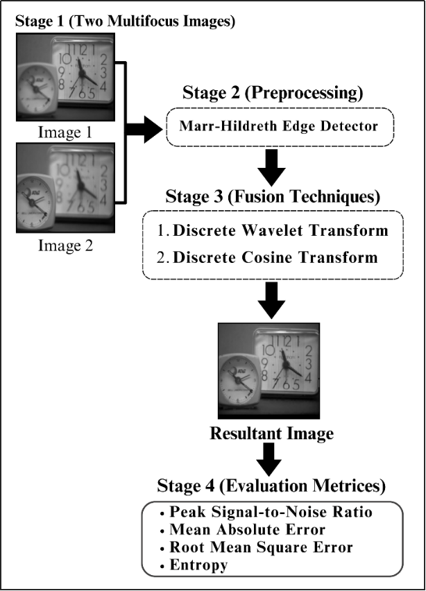
Pre-processing in image fusion refers to processes and methods applied to input images before these images are combined or merged into one for a single, enhanced image with higher visual quality and information content [20]. The objective of image fusion pre-processing is to prepare input images for effective fusion, where the most important and usable information will be contained in the result of the fused image [21]. A few of the techniques of pre-processing that the framework uses are described:
The Marr–Hildreth technique is a widely used edge detection method in image processing that enhances the clarity of object boundaries by detecting regions of rapid intensity change. It applies Gaussian smoothing to reduce noise and then computes the Laplacian to identify zero-crossings, which represent edges in the image [22]. This approach effectively highlights fine details and structural features, making it particularly useful for applications requiring precise edge detection, such as medical imaging, remote sensing, and object recognition [23]. In the context of multi-focus image fusion, the Marr–Hildreth technique helps refine image details before fusion, ensuring that only the most relevant and well-defined features contribute to the final composite image [24]. Although Marr–Hildreth Edge Detection (MHED) is traditionally utilized to detect edges in an image, it is adapted here as part of the preprocessing step. Instead of using it for edge detection, MHED is employed to enhance the overall sharpness and edge clarity of the input images. This enhancement enables the fusion algorithms to better combine the information from multiple sources, resulting in a higher-quality fused image. The modified MHED process prepares the images by sharpening them and making edge details more prominent, which is essential for effective fusion.
After pre-processing, fusion combines enhanced images into a single, informative representation. This amalgamation enhances visual quality, detail richness, and overall scene representation. Fusion maximizes improvement potential by implementing diverse enhancement techniques, resulting in a final image that effectively communicates desired information. Techniques include:
The Stationary Wavelet Transform (SWT) is an advanced multi-resolution analysis technique used in image processing to enhance and preserve important image details. Unlike the traditional Discrete Wavelet Transform (DWT), SWT is shift-invariant, meaning it does not suffer from information loss due to down-sampling. This property makes SWT particularly useful for applications such as image denoising, enhancement, and fusion, where maintaining spatial consistency is crucial. In multi-focus image fusion, SWT effectively decomposes images into different frequency components while retaining spatial alignment, ensuring that critical details from each source image are accurately preserved and combined into a single, sharp, and well-focused composite image [25].
This approach applies wavelets to get uncertain signals because of noise, and it has proven very useful for processing X-ray images, magnetic resonance images, and applications in medical science. In this approach, it is possible to enhance the image without distortion or blurring of finer features. The DWT applied in this method gives adaptive spatial frequency resolution; that is, it improves the frequency resolution at low frequencies and enhances the spatial resolution at high frequencies. The DWT does this compactly by representing the frequency components of the signal so that the resolution is in real-time and gives better data about frequency. A better image is generated using DWT through non-duplicative graphic representation features, which furtherimproves the spectral and spatial localization [25]. Based on the concept of multiresolution analysis, a fast technique under the title of Wavelet Transform was developed; it tends to have both temporal and frequency discrete properties. The Wavelet Transform has the property of providing a complete representation by covering both time and frequency features in an image quickly. This method decomposes the data into two sets: high-frequency and low-frequency components. Decomposition is the division process that helps to derive statistics related to low and high frequencies [26].
The wavelet transform has been one of the important logical tools developed for describing images at different resolutions. The basic idea behind what has made wavelet transform theory possible is the mother wavelet. This idea of the mother wavelet has found applications, and many more are likely to be realized as technology advances in texture analysis, image compression, digital image watermarking, and facial recognition. The classification of wavelets comes under two kinds: continuous and discrete. This new tool influences many analytical domains, not necessarily image processing [27]. The discrete wavelet transforms (DWT) use wavelet coefficients, scale factors, and functions. The DWT may be represented as a discrete signal or image, , as shown by Eq. 1:
in Eq. 1 are wavelet coefficients at scale and translation . is the input signal or image. is the symbol of the wavelet function. The frequency resolution is determined by the scale factor denoted by . A factor defines the position of the wavelet function and, in turn, is a translation or shift factor.
The DWT shows the input signal or image in different levels of expressions, which is done in an iterative process by applying this expression to the decomposition layers. The Wavelet Transform is a function receiving two inputs. When the scale increases, the signal's peaks become more similar, but greater translation values are necessary for the tails of the signal to resemble one another [28]. The Discrete Wavelet Transform can extract hidden changes by analysing signals and visuals from a measurable time-frequency point of view. Wavelets explore sharp changes in signals or visual [29]. In this wavelet transform method, the signal isprojected onto a set of wavelet functions, which decomposes the signal into a sequence of wavelet functions. To put it another way, this transformation is a complete conversion [30].
The Discrete Wavelet Transform (DWT) includes advantages such as analyzing signals and images at various sizes and capturing localized and global information. DWT also offers multiscale analysis, effective data compression, feature extraction, and pattern recognition. One of the downsides of the Discrete Wavelet Transform (DWT) is that the fused pictures lose responsiveness to directional features [31].
Discrete Cosine Transform (DCT) image fusion method is commonly used to fuse images because of its effectiveness and good properties. The utilization of DCT is needed to transform the original images into frequency domain followed by fusing the obtained DCT coefficients. Finally, the parameters are synthesized in spatial domain to obtain the fused image. One of the good properties of DCT-based fusion methods is their capability to keep high frequency information, which is important for retaining image features [32]. While it is an important transformation in various problems of science and engineering, the key thing where this concept is needed for our focus is for picture compression. DCT offers an efficient transformation, converting a picture from the spatial to frequency domain increasing its applicative area [33]. The overall framework of the proposed multi-focus image fusion method is illustrated in Figure 2, highlighting the main components and processing pipeline.
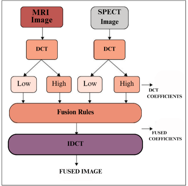
DCT is known for its computational efficiency and low memory consumption, which makes it better for image fusion applications. DCT-based fusion has a significant drawback: its sensitivity to image misalignment. When the source images aren't properly aligned, the fused image can exhibit undesirable artifacts such as ringing or blurring. For this purpose, DCT based fusion combined with image fusion techniques can be used. Before carrying out fusion, it is therefore critical to register the source images as perfectly aligned results in a marked reduction of artefacts and hence significantly improved precision. This results in outputs of higher quality when the strengths and weaknesses or DCT-based fusion are combined properly [32].
This research experiment is done on Core i5 10th generation system with a ram of 16GB which uses MATLAB for implementation. MATLAB tool was employed for pre-processing, image fusion and performance evaluation.
In this research experiment, we used four different sets of grayscales multifocus images. Figure 1 shows the Lytro Multifocus dataset [20]. This dataset is popular among researchers worldwide for multifocus image experiments. It includes images of clocks, balloons, leaves, and bottles.
The performance measurements employed in this study included entropy, peak signal-to-noise ratio (PSNR), mean absolute error (MAE) and root mean square error. Without these metrices, it is impossible to calculate the weight of how effectively those techniques are working in an unbiased way. These are the widely accepted metrices that gives a formal and systematic way for evaluating performance and efficiency of novel techniques.
RMSE is frequently used to compare the pixel differences between original and generated images. It provides a good measure of the final image's quality by indicating how accurately the colours and brightness are represented [34]. Equation 1 shows the RMSE formula.
The Peak Signal-to-Noise Ratio (PSNR) measures the quality of an image by comparing the original and fused images. It measures how similar the pixels are by comparing the peak signal strength to the noise by comparing the number of matching pixels to the total number of gray levels. Equation 2 shows PSNR formula.
This section presents the results of all the research experiments carried out in this study. We performed both qualitative and quantitative evaluation of various image fusion and enhancement techniques to provide a comprehensive interpretation of the results.
The proposed methodology was tested on different datasets such as clocks, balloons, leaves, and bottles. The results achieved by the proposed method were compared withimage fusion techniques using the same datasets. To provide a comprehensive comparison, the results were analyzed both with and without the application of the Marr–Hildreth technique for image enhancement.Both quantitative and quantitative methodologies were used to compare the results with the proposed novel framework. This maingoal of this extensive testing is to evaluate the methodology's effectiveness for multi-focus images. For quantitative analysis, evaluation metrices such as PSNR, MAE, RMSE and entropy are used in this study. All of these metrices led to a clear view of the level of the fused images that came from the proposed methodology. Furthermore, the qualitative analysis allowed visual checking of the changes, improvements, and corrections made in the fused images. The results of these performance measures will now provide a clear insight into the functionality and effectiveness of the applied image fusion techniques of the present study. The output has clearly shown benefits and efficiency for the suggested methodology in image fusion.
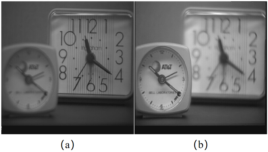
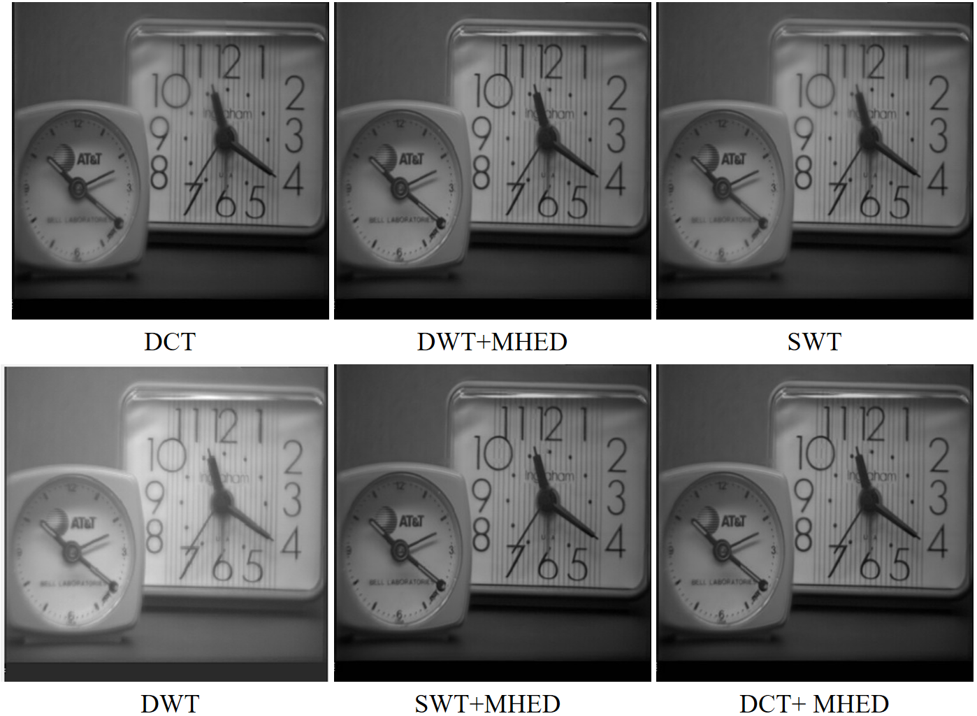
Figure 3 shows a set of two multifocus images, where (a) and (b) represent the original images used for the study. The fused enhanced images in Figure 4 were created by combining them using a variety of methods, including DWT, SWT, DCT, DCT+MHED, DWT+MHED, and SWT+MHED.
| Fusion Techniques | PSNR | RMSE | MAE | E |
|---|---|---|---|---|
| DWT | 39.6314 | 2.6605 | 24.8771 | 7.4158 |
| SWT | 38.5665 | 3.0075 | 28.6275 | 7.4215 |
| DWT+MHED | 39.7077 | 2.6372 | 17.2962 | 7.4311 |
| DCT | 39.6266 | 2.6620 | 24.8633 | 7.4155 |
| SWT+MHED | 39.7074 | 2.6373 | 17.2950 | 7.4313 |
| DCT+MHED | 39.7990 | 2.6302 | 17.2633 | 7.4320 |
Table 2 compares different multi-focus image fusion methods using the Clock and Leaves image set, based on PSNR, RMSE, MAE, and entropy (E). The DCT+MHED method achieved the highest PSNR of 39.7990, meaning it produced the clearest and most detailed image. It also had the lowest MAE of 17.2633, showing minimal error, and the highest entropy of 7.4320, indicating better detail preservation. Similarly, DWT+MHED and SWT+MHED also performed well, proving that adding the Marr–Hildreth Edge Detection (MHED) technique improves the overall fusion quality.
In contrast, the SWT method had the lowest PSNR of 38.5665 and the highest MAE of 28.6275, meaning it did not preserve details as effectively. Likewise, DWT and DCT without enhancement had higher RMSE values of 2.6605 and 2.6620, indicating more errors. These results clearly show that using enhancement techniques like MHED helps create sharper and more detailed fused images.
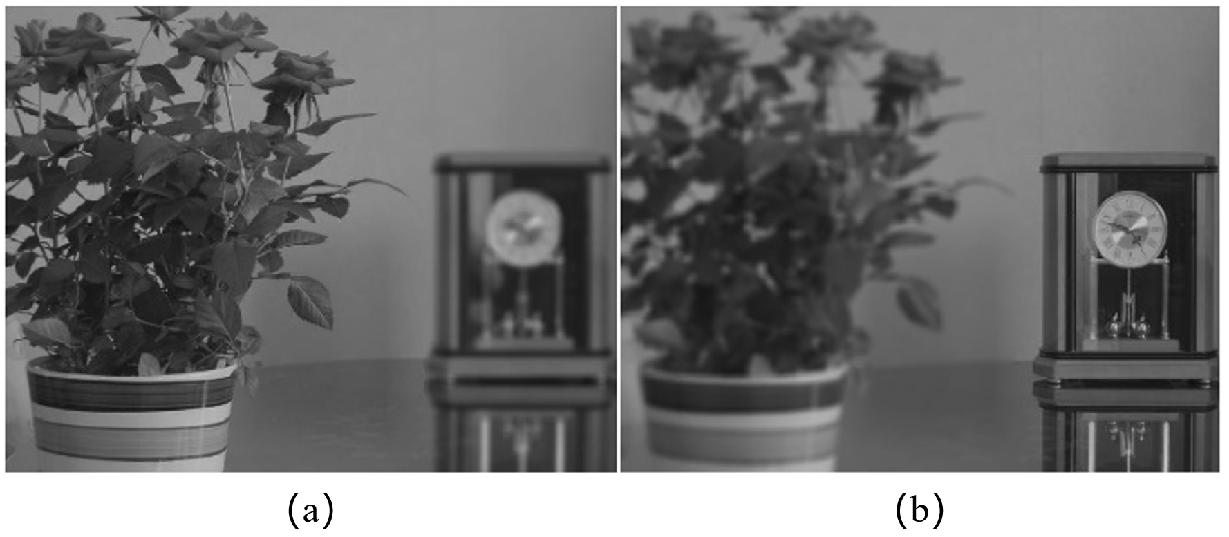
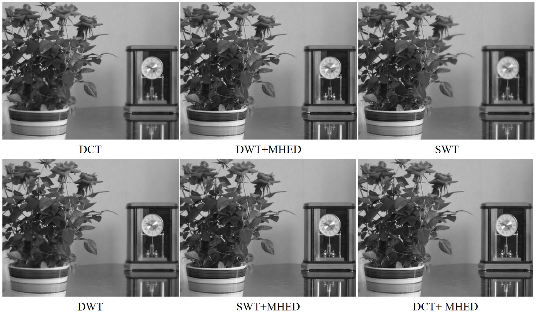
Figure 5 shows a set of two multifocus images, where (a) and (b) represent the original images used for the study. The fused enhanced images in Figure 6 were created by combining them using a variety of methods, including DWT, SWT, DCT, DCT+MHED, DWT+MHED, and SWT+MHED.
| Fusion Techniques | PSNR | RMSE | MAE | E |
|---|---|---|---|---|
| DWT | 40.7087 | 2.3501 | 19.4622 | 6.9863 |
| SWT | 41.4077 | 2.1682 | 18.9728 | 6.9942 |
| DWT+MHED | 39.7810 | 2.6150 | 15.7361 | 7.0041 |
| DCT | 41.4014 | 2.1700 | 18.9641 | 6.9898 |
| SWT + MHED | 39.7828 | 2.6145 | 15.7375 | 7.0085 |
| DCT+MHED | 39.7724 | 2.6176 | 15.7114 | 7.0041 |
Table 3 presents the results of different multi-focus image fusion methods using the Clocks image set, evaluated using PSNR, RMSE, MAE, and entropy (E). The SWT method achieved the highest PSNR of 41.4077, indicating that it produced the sharpest and most detailed image. Similarly, DCT closely followed with a PSNR of 41.4014, showing that it effectively preserved image quality. However, the addition of MHED (Marr–Hildreth Edge Detection) in DWT+MHED, SWT+MHED, and DCT+MHED resulted in improved entropy values, highlighting better detail retention.
On the other hand, DWT+MHED and DCT+MHED showed lower MAE values of 15.7361 and 15.7114, respectively, indicating fewer errors and better accuracy. However, DWT alone had a relatively lower performance with a PSNR of 40.7087 and a higher MAE of 19.4622, suggesting that without enhancement techniques, it may not retain fine details as effectively. These results emphasize that applying enhancement techniques like MHED improves image fusion quality by refining details and reducing errors.
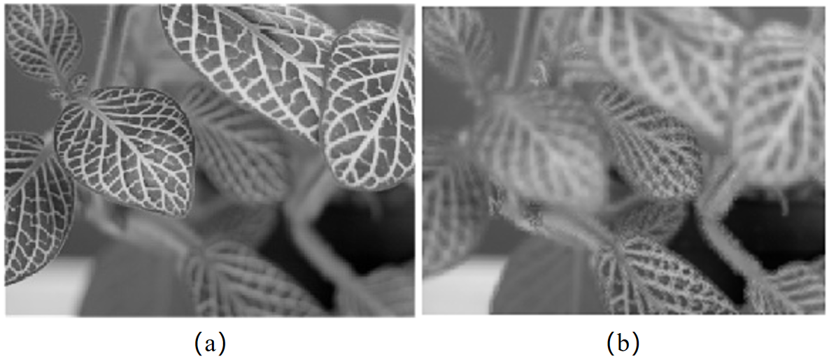
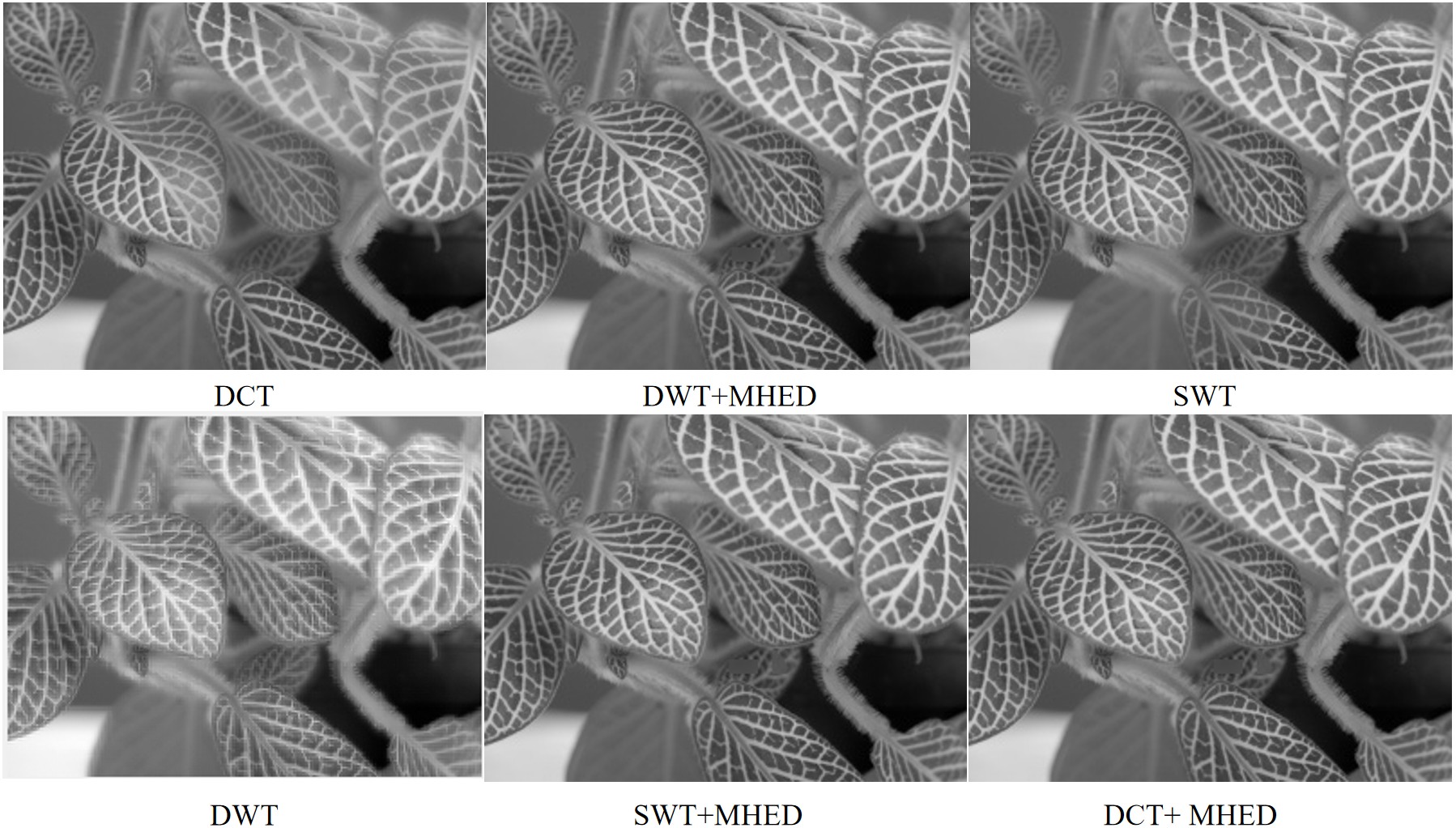
Figure 7 shows a set of two multifocus images, where (a) and (b) represent the original images used for the study. The fused enhanced images in Figure 8 were created by combining them using a variety of methods, including DWT, SWT, DCT, DCT+MHED, DWT+MHED, and SWT+MHED.
| Fusion Techniques | PSNR | RMSE | MAE | E |
|---|---|---|---|---|
| DWT | 33.7621 | 5.2291 | 43.3788 | 7.2993 |
| SWT | 38.2289 | 3.1267 | 23.2605 | 7.3099 |
| DWT+MHED | 40.2055 | 7.3447 | 8.7347 | 7.3447 |
| DCT | 33.7615 | 5.2295 | 43.3852 | 7.2999 |
| SWT + MHED | 40.2078 | 2.4896 | 8.7319 | 7.3447 |
| DCT+MHED | 40.1937 | 2.4937 | 8.7438 | 7.3454 |
Table 4 presents the performance of different multi-focus image fusion methods using the Leaves image set, evaluated using PSNR, RMSE, MAE, and entropy (E). The SWT+MHED method achieved the highest PSNR of 40.2078, indicating superior image quality with well-preserved details. Similarly, DWT+MHED and DCT+MHED produced high PSNR values above 40, demonstrating that applying Marr–Hildreth Edge Detection (MHED) significantly improved image sharpness and clarity.
On the other hand, DWT and DCT without enhancement yielded lower PSNR values of 33.7621 and 33.7615, respectively, along with higher MAE values of over 43, indicating weaker detail preservation. The SWT method alone also performed well, with a PSNR of 38.2289, but integrating MHED further reduced error and enhanced fine details.
Overall, these results highlight that using enhancement techniques like MHED in combination with transformation methods leads to better fusion quality by minimizing errors and improving edge definition.
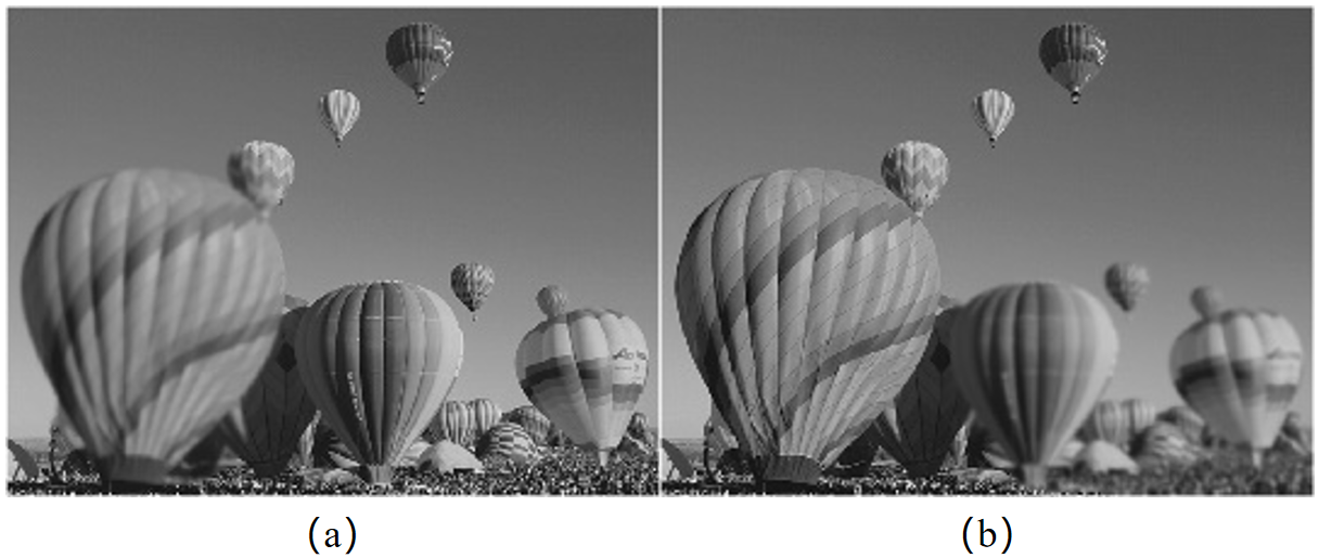
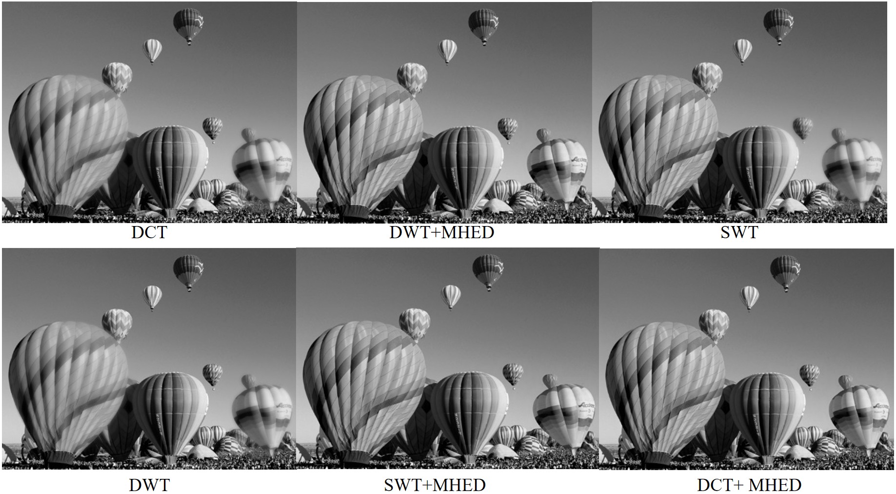
Figure 9 shows a set of two multifocus images, where (a) and (b) represent the original images used for the study. The fused enhanced images in Figure 10 were created by combining them using a variety of methods, including DWT, SWT, DCT, DCT+MHED, DWT+MHED, and SWT+MHED.
| Fusion Techniques | PSNR | RMSE | MAE | E |
|---|---|---|---|---|
| DWT | 37.5477 | 3.3818 | 31.7169 | 7.4465 |
| SWT | 41.1553 | 2.2834 | 12.2743 | 7.1480 |
| DWT+ MHED | 41.3035 | 2.1946 | 9.9225 | 7.4472 |
| DCT | 37.5437 | 3.3833 | 31.71515 | 7.4474 |
| SWT + MHED | 41.3010 | 2.1952 | 9.9227 | 7.4482 |
| DCT+MHED | 41.2940 | 2.1970 | 9.9274 | 7.4498 |
Table 5 presents the results of different multi-focus image fusion techniques applied to the Balloons image set, evaluated using PSNR, RMSE, MAE, and entropy (E). The DWT+MHED method achieved the highest PSNR of 41.3035, indicating superior image quality with well-preserved details. Similarly, SWT+MHED and DCT+MHED also performed well, achieving PSNR values above 41, demonstrating that integrating Marr–Hildreth Edge Detection (MHED) significantly improved sharpness and clarity.
On the other hand, DWT and DCT without enhancement yielded lower PSNR values of 37.5477 and 37.5437, respectively, with higher MAE values exceeding 31, suggesting weaker edge preservation. The SWT method alone also performed well, but incorporating MHED further reduced errors and enhanced fine details.
These results highlight the effectiveness of combining transformation techniques with edge enhancement methods like MHED, which significantly improve the quality of fused images by reducing errors and enhancing clarity.
This paper presents a significant development in image processing, demonstrating the advantages of integrating multi-focus image fusion techniques based on the Marr–Hildreth technique, DCT, SWT, and DWT. The resulting clarity and detail can enhance results in medical imaging, remote sensing, and photography. The structured methodology provides a reliable way to produce high-quality fused images, providing further analysis and visualization. These findings lay a foundation for future studies to refine and expand these methods, potentially making a pioneering contribution to image fusion technology.
This work demonstrates the performance of versatile image fusion technology based on the Marr–Hildreth technique, DCT, SWT, and DWT with enhancement and without prior masking. It is limited to grayscale images from the Lytro dataset. The application to other types of images or datasets has not been explored. Additionally, while DCT, SWT, and DWT have shown promising results, other pre-processing and fusion methods were not investigated in this research. The experiments were conducted in a controlled MATLAB environment, which may differ from real-world applications. Future research could expand on these areas to further validate and enhance the findings.
This research study suggested a novel technique for multi-focus image fusion using the Marr–Hildreth technique, Discrete Cosine Transform, Stationary Wavelet Transform, and Discrete Wavelet Transform. The experimental results show that both qualitative and quantitative evaluations have significantly improved. Furthermore, when applied to the multi-focus image datasets, the proposed technique works well. The results are significant for image fusion and enhancement in the field, revealing the multiplicity of ways researchers approach solving multi-focus image challenges. It also provides a better understanding of how to develop image fusion and processing research in the new horizon of advanced scientific investigation, an emerging domain.
The research limitations are that the study focuses only on grayscale images from the Lytro dataset using DCT, SWT, and DWT-based multi-focus image fusion techniques in MATLAB, without considering other types of images, datasets, or different pre-processing and fusion methods. Future work will expand on this by testing the proposed techniques on a broader range of image types and datasets, including color images. Additionally, exploring other pre-processing and fusion methods could further enhance image quality. Implementing and evaluating these techniques in real-world scenarios would help validate their practical applications. Integrating advanced machine learning algorithms could also offer new ways to optimize the image fusion process, leading to greater improvements in clarity and detail. In conclusion, while Marr-Hildreth Edge Detection (MHED) is typically used for edge detection, in this study, it served a unique role as a pre-processing step. Its primary function was to enhance the sharpness and clarity of the input images before fusion, which proved crucial in improving the overall quality of the fused images. This demonstrates the versatility of MHED in image processing beyond its traditional use.
 Copyright © 2025 by the Author(s). Published by Institute of Central Computation and Knowledge. This article is an open access article distributed under the terms and conditions of the Creative Commons Attribution (CC BY) license (https://creativecommons.org/licenses/by/4.0/), which permits use, sharing, adaptation, distribution and reproduction in any medium or format, as long as you give appropriate credit to the original author(s) and the source, provide a link to the Creative Commons licence, and indicate if changes were made.
Portico
All published articles are preserved here permanently:
https://www.portico.org/publishers/icck/