

Magnetic Resonance Imaging Findings of Unilateral Temporomandibular Joint Derangement and Condylar Morphological Changes in a 14-Year-Old Patient: A Case Report



Article Information
Abstract
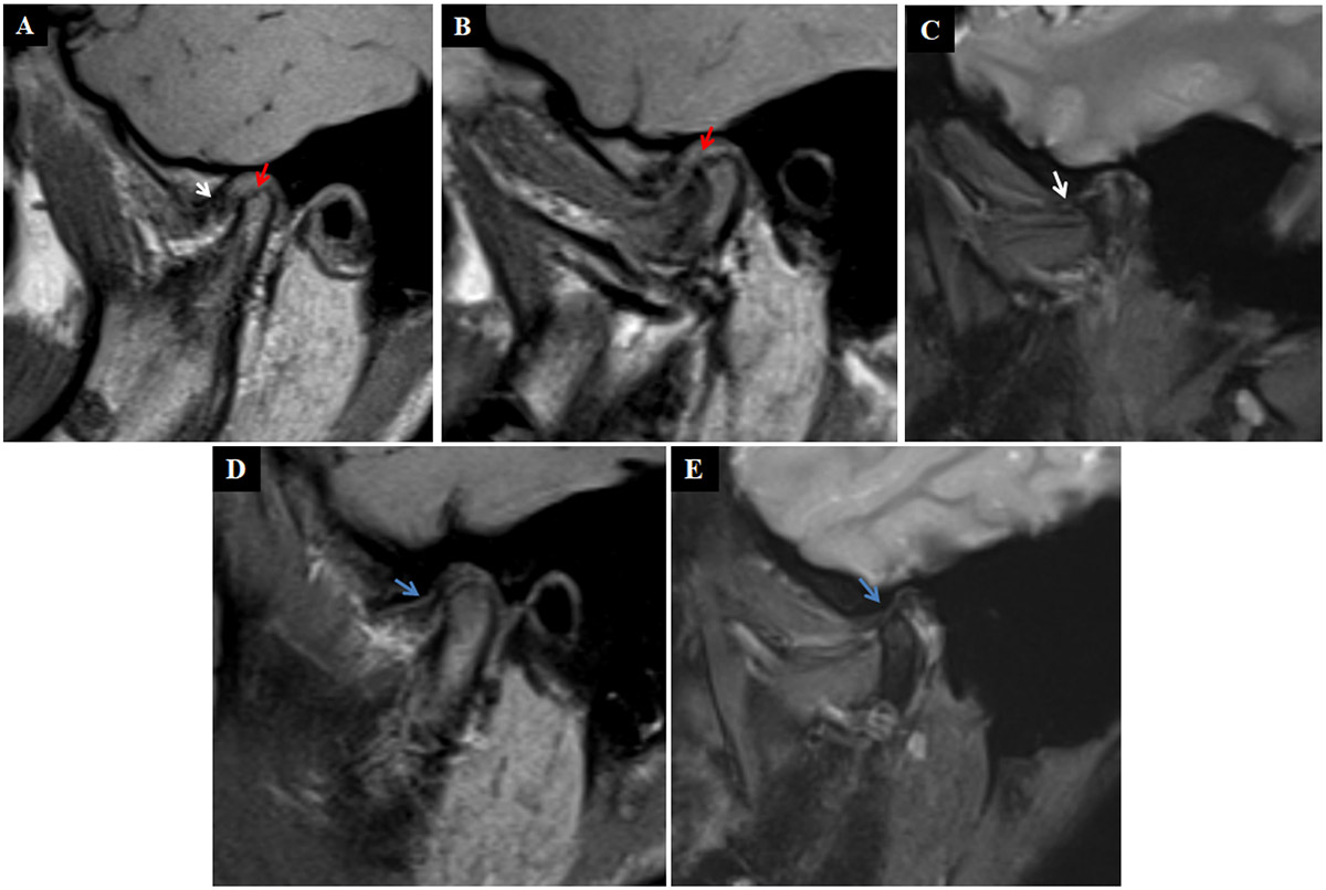
Temporomandibular disorders may occur during adolescence, although the relationship between clinical symptoms and structural joint abnormalities remains incompletely understood. Magnetic resonance imaging (MRI) allows detailed assessment of articular disc position and morphology and of the disc--condyle relationship. This report describes unilateral discal and condylar abnormalities in a symptomatic adolescent. A 14-year-old female presented with a one-year history of right-sided preauricular pain, joint clicking, and restricted mouth opening. She also reported sleep bruxism and childhood jaw trauma. Bilateral temporomandibular joint (TMJ) MRI showed anterior displacement of the right articular disc without reduction, with irregular disc morphology; the disc remained anteriorly displaced in the open-mouth position. Flattening and irregularity of the right condylar articular surface were observed, together with increased thickness and signal intensity of the posterior disc attachment and minimal intra-articular fluid. The left TMJ showed preserved disc morphology, condylar contour, and a normal disc--condyle relationship in both mouth positions, with a minimal amount of intra-articular fluid. This case documents the coexistence of persistent unilateral TMJ symptoms with discal and condylar abnormalities during adolescence, and bilateral closed- and open-mouth MRI clarified the distribution and extent of intra-articular involvement. Although a single examination cannot establish the relationship between disc displacement and condylar changes, the findings support comprehensive clinical assessment and individualized follow-up in adolescents with persistent TMJ symptoms.
Graphical Abstract
Keywords
Data Availability Statement
Funding
Conflicts of Interest
AI Use Statement
Ethical Approval and Consent to Participate
References
- Li, D. T. S., & Leung, Y. Y. (2021). Temporomandibular disorders: Current concepts and controversies in diagnosis and management. Diagnostics, 11(3), 459.
[CrossRef] [Google Scholar] - Rauch, A., Schierz, O., Körner, A., Kiess, W., & Hirsch, C. (2020). Prevalence of anamnestic symptoms and clinical signs of temporomandibular disorders in adolescents—Results of the epidemiologic LIFE Child Study. Journal of Oral Rehabilitation, 47(4), 425-431.
[CrossRef] [Google Scholar] - Rongo, R., Ekberg, E., Nilsson, I. M., Al‐Khotani, A., Alstergren, P., Conti, P. C. R., ... & Michelotti, A. (2021). Diagnostic criteria for temporomandibular disorders (DC/TMD) for children and adolescents: An international Delphi study—Part 1‐Development of Axis I. Journal of Oral Rehabilitation, 48(7), 836-845.
[CrossRef] [Google Scholar] - Tominaga, K., Konoo, T., Morimoto, Y., Tanaka, T., Habu, M., & Fukuda, J. (2007). Changes in temporomandibular disc position during growth in young Japanese. Dentomaxillofacial Radiology, 36(7), 397-401.
[CrossRef] [Google Scholar] - Li, C., & Zhang, Q. (2023). Comparison of imaging findings of 714 symptomatic and asymptomatic temporomandibular joints: a retrospective study. BMC Oral Health, 23(1), 79.
[CrossRef] [Google Scholar] - Mallya, S. M., Ahmad, M., Cohen, J. R., Kaspo, G., & Ramesh, A. (2022). Recommendations for imaging of the temporomandibular joint. Position statement from the American Academy of Oral and Maxillofacial Radiology and the American Academy of Orofacial Pain. Oral Surgery, Oral Medicine, Oral Pathology and Oral Radiology, 134(5), 639-648.
[CrossRef] [Google Scholar] - De Stefano, A. A., Guercio-Mónaco, E., Hernández-Andara, A., Horodynski, M., & Galluccio, G. (2025). Association Between Temporomandibular Disk Position and Condylar and Fossa Morphology in Adolescents: A Cross-Sectional Study. FACE, 6(3), 456-465.
[CrossRef] [Google Scholar] - Kumar, R., Pallagatti, S., Sheikh, S., Mittal, A., Gupta, D., & Gupta, S. (2015). Correlation between clinical findings of temporomandibular disorders and MRI characteristics of disc displacement. The Open Dentistry Journal, 9, 273-281.
[CrossRef] [Google Scholar] - Li, C. X., Liu, X., Gong, Z. C., Jumatai, S., & Ling, B. (2022). Morphologic analysis of condyle among different disc status in the temporomandibular joints by three-dimensional reconstructive imaging: a preliminary study. BMC Oral Health, 22(1), 395.
[CrossRef] [Google Scholar] - Junhasavasdikul, T., Abadeh, A., Tolend, M., & Doria, A. S. (2018). Developing a reference MRI database for temporomandibular joints in healthy children and adolescents. Pediatric Radiology, 48(8), 1113--1122.
[CrossRef] [Google Scholar] - Wu, S., Zhang, D., Xia, S., Shen, P., & Yang, C. (2025). Effect of temporomandibular joint anterior disc displacement on condylar height in different age groups. BMC Oral Health, 25(1), 1100.
[CrossRef] [Google Scholar] - Bottini, G. B., Roccia, F., & Sobrero, F. (2024). Management of pediatric mandibular condyle fractures: A literature review. Journal of Clinical Medicine, 13(22), 6921.
[CrossRef] [Google Scholar] - Topaloglu-Ak, A., Kurtulmus, H., Basa, S., & Sabuncuoglu, O. (2022). Can sleeping habits be associated with sleep bruxism, temporomandibular disorders and dental caries among children? Dental and medical problems, 59(4), 517-522.
[CrossRef] [Google Scholar] - Meto, A., & Halilaj, G. (2025). The integration of cone beam computed tomography, artificial intelligence, augmented reality, and virtual reality in dental diagnostics, surgical planning, and education: A narrative review. Applied Sciences, 15(11), 6308.
[CrossRef] [Google Scholar]
Cite This Article
TY  - JOUR
AU  - Halilaj, Gerta
AU  - Stroni, Gentian
PY  - 2026
DA  - 2026/11/11
TI  - Magnetic Resonance Imaging Findings of Unilateral Temporomandibular Joint Derangement and Condylar Morphological Changes in a 14-Year-Old Patient: A Case Report
JO  - Journal of Oral and Dental Approaches
T2  - Journal of Oral and Dental Approaches
JF  - Journal of Oral and Dental Approaches
VL  - 1
IS  - 1
SP  - 13
EP  - 19
DO  - 10.62762/JODA.2026.471521
UR  - https://www.icck.org/article/abs/joda.2026.471521
KW  - adolescent
KW  - temporomandibular joint
KW  - magnetic resonance imaging
KW  - articular disc displacement
AB  - Temporomandibular disorders may occur during adolescence, although the relationship between clinical symptoms and structural joint abnormalities remains incompletely understood. Magnetic resonance imaging (MRI) allows detailed assessment of articular disc position and morphology and of the disc--condyle relationship. This report describes unilateral discal and condylar abnormalities in a symptomatic adolescent. A 14-year-old female presented with a one-year history of right-sided preauricular pain, joint clicking, and restricted mouth opening. She also reported sleep bruxism and childhood jaw trauma. Bilateral temporomandibular joint (TMJ) MRI showed anterior displacement of the right articular disc without reduction, with irregular disc morphology; the disc remained anteriorly displaced in the open-mouth position. Flattening and irregularity of the right condylar articular surface were observed, together with increased thickness and signal intensity of the posterior disc attachment and minimal intra-articular fluid. The left TMJ showed preserved disc morphology, condylar contour, and a normal disc--condyle relationship in both mouth positions, with a minimal amount of intra-articular fluid. This case documents the coexistence of persistent unilateral TMJ symptoms with discal and condylar abnormalities during adolescence, and bilateral closed- and open-mouth MRI clarified the distribution and extent of intra-articular involvement. Although a single examination cannot establish the relationship between disc displacement and condylar changes, the findings support comprehensive clinical assessment and individualized follow-up in adolescents with persistent TMJ symptoms.
SN  - pending
PB  - Institute of Central Computation and Knowledge
LA  - English
ER  -
@article{Halilaj2026Magnetic,
  author = {Gerta Halilaj and Gentian Stroni},
  title = {Magnetic Resonance Imaging Findings of Unilateral Temporomandibular Joint Derangement and Condylar Morphological Changes in a 14-Year-Old Patient: A Case Report},
  journal = {Journal of Oral and Dental Approaches},
  year = {2026},
  volume = {1},
  number = {1},
  pages = {13-19},
  doi = {10.62762/JODA.2026.471521},
  url = {https://www.icck.org/article/abs/joda.2026.471521},
  abstract = {Temporomandibular disorders may occur during adolescence, although the relationship between clinical symptoms and structural joint abnormalities remains incompletely understood. Magnetic resonance imaging (MRI) allows detailed assessment of articular disc position and morphology and of the disc--condyle relationship. This report describes unilateral discal and condylar abnormalities in a symptomatic adolescent. A 14-year-old female presented with a one-year history of right-sided preauricular pain, joint clicking, and restricted mouth opening. She also reported sleep bruxism and childhood jaw trauma. Bilateral temporomandibular joint (TMJ) MRI showed anterior displacement of the right articular disc without reduction, with irregular disc morphology; the disc remained anteriorly displaced in the open-mouth position. Flattening and irregularity of the right condylar articular surface were observed, together with increased thickness and signal intensity of the posterior disc attachment and minimal intra-articular fluid. The left TMJ showed preserved disc morphology, condylar contour, and a normal disc--condyle relationship in both mouth positions, with a minimal amount of intra-articular fluid. This case documents the coexistence of persistent unilateral TMJ symptoms with discal and condylar abnormalities during adolescence, and bilateral closed- and open-mouth MRI clarified the distribution and extent of intra-articular involvement. Although a single examination cannot establish the relationship between disc displacement and condylar changes, the findings support comprehensive clinical assessment and individualized follow-up in adolescents with persistent TMJ symptoms.},
  keywords = {adolescent, temporomandibular joint, magnetic resonance imaging, articular disc displacement},
  issn = {pending},
  publisher = {Institute of Central Computation and Knowledge}
}
Article Metrics
Publisher's Note
ICCK stays neutral with regard to jurisdictional claims in published maps and institutional affiliations.
Rights and Permissions
 Copyright © 2026 by the Author(s). Published by Institute of Central Computation and Knowledge. This article is an open access article distributed under the terms and conditions of the Creative Commons Attribution (CC BY) license (https://creativecommons.org/licenses/by/4.0/), which permits use, sharing, adaptation, distribution and reproduction in any medium or format, as long as you give appropriate credit to the original author(s) and the source, provide a link to the Creative Commons licence, and indicate if changes were made.