


A Tri-Phasic View of the Microenvironment in Breast Cancer Brain Metastasis



Article Information
Abstract
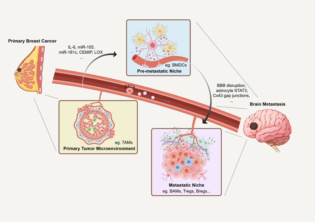
Breast cancer brain metastasis (BCBM) is driven by microenvironmental remodeling across three interconnected phases: the primary tumor microenvironment (TME) licenses dissemination, the pre-metastatic niche (PMN) primes the brain, and the established metastatic niche sustains outgrowth through central nervous system (CNS)-resident cell interactions. These phases are individually established; we integrate them through cross-phase signaling axes, exemplified by the proposed IL-6$\rightarrow$extracellular vesicle (EV)$\rightarrow$blood-brain barrier (BBB) disruption cascade, that link tumor-associated macrophage-driven epithelial-mesenchymal transition to PMN conditioning and niche maintenance. We integrate single-cell and spatial transcriptomic insights, discuss brain-specific mechanisms including astrocytic STAT3/connexin 43 signaling, neuronal contributions, and the neurovascular unit, and evaluate emerging therapies with emphasis on antibody-drug conjugates with intracranial activity. Subtype-specific differences between HER2-positive and triple-negative breast cancer brain metastases and key translational barriers are systematically addressed.
Graphical Abstract
Keywords
Data Availability Statement
Funding
Conflicts of Interest
AI Use Statement
Ethical Approval and Consent to Participate
References
- Giaquinto, A. N., Sung, H., Newman, L. A., Freedman, R. A., Smith, R. A., Star, J., ... & Siegel, R. L. (2024). Breast cancer statistics 2024. CA: a cancer journal for clinicians, 74(6), 477-495.
[CrossRef] [Google Scholar] - Kuksis, M., Gao, Y., Tran, W., Hoey, C., Kiss, A., Komorowski, A. S., ... & Jerzak, K. J. (2021). The incidence of brain metastases among patients with metastatic breast cancer: a systematic review and meta-analysis. Neuro-oncology, 23(6), 894-904.
[CrossRef] [Google Scholar] - Priego, N., Zhu, L., Monteiro, C., Mulders, M., Wasilewski, D., Bindeman, W., ... & Valiente, M. (2018). STAT3 labels a subpopulation of reactive astrocytes required for brain metastasis. Nature medicine, 24(7), 1024-1035.
[CrossRef] [Google Scholar] - Zhou, W., Fong, M. Y., Min, Y., Somlo, G., Liu, L., Palomares, M. R., ... & Wang, S. E. (2014). Cancer-secreted miR-105 destroys vascular endothelial barriers to promote metastasis. Cancer cell, 25(4), 501-515.
[CrossRef] [Google Scholar] - Tominaga, N., Kosaka, N., Ono, M., Katsuda, T., Yoshioka, Y., Tamura, K., ... & Ochiya, T. (2015). Brain metastatic cancer cells release microRNA-181c-containing extracellular vesicles capable of destructing blood–brain barrier. Nature communications, 6(1), 6716.
[CrossRef] [Google Scholar] - Erler, J. T., Bennewith, K. L., Cox, T. R., Lang, G., Bird, D., Koong, A., ... & Giaccia, A. J. (2009). Hypoxia-induced lysyl oxidase is a critical mediator of bone marrow cell recruitment to form the premetastatic niche. Cancer cell, 15(1), 35-44.
[CrossRef] [Google Scholar] - Rodrigues, G., Hoshino, A., Kenific, C. M., Matei, I. R., Steiner, L., Freitas, D., ... & Lyden, D. (2019). Tumour exosomal CEMIP protein promotes cancer cell colonization in brain metastasis. Nature cell biology, 21(11), 1403-1412.
[CrossRef] [Google Scholar] - Gong, X., Hou, Z., Endsley, M. P., Gronseth, E. I., Rarick, K. R., Jorns, J. M., \ldots, & Wang, L. (2019). Interaction of tumor cells and astrocytes promotes breast cancer brain metastases through TGF-$\beta$2/ANGPTL4 axes. NPJ Precision Oncology, 3(1), 24. \href{
[CrossRef] [Google Scholar] - Fong, M. Y., Zhou, W., Liu, L., Alontaga, A. Y., Chandra, M., Ashby, J., ... & Wang, S. E. (2015). Breast-cancer-secreted miR-122 reprograms glucose metabolism in premetastatic niche to promote metastasis. Nature cell biology, 17(2), 183-194.
[CrossRef] [Google Scholar] - Lamba, N., Wen, P. Y., & Aizer, A. A. (2021). Epidemiology of brain metastases and leptomeningeal disease. Neuro-oncology, 23(9), 1447-1456.
[CrossRef] [Google Scholar] - Arvanitis, C. D., Ferraro, G. B., & Jain, R. K. (2020). The blood–brain barrier and blood–tumour barrier in brain tumours and metastases. Nature Reviews Cancer, 20(1), 26-41.
[CrossRef] [Google Scholar] - Dongre, A., Rashidian, M., Reinhardt, F., Bagnato, A., Keckesova, Z., Ploegh, H. L., & Weinberg, R. A. (2017). Epithelial-to-mesenchymal transition contributes to immunosuppression in breast carcinomas. Cancer research, 77(15), 3982-3989.
[CrossRef] [Google Scholar] - Qi, Y., Li, R., & Han, M. (2024). Tumor-associated macrophages induce epithelial-mesenchymal transition and promote lung metastasis in breast cancer by activating the IL-6/STAT3/TGM2 axis. Int Immunopharmacol, 143(Pt 2), 113387.
[CrossRef] [Google Scholar] - Becker, A., Thakur, B. K., Weiss, J. M., Kim, H. S., Peinado, H., & Lyden, D. (2016). Extracellular vesicles in cancer: cell-to-cell mediators of metastasis. Cancer cell, 30(6), 836-848.
[CrossRef] [Google Scholar] - Peinado, H., Zhang, H., Matei, I. R., Costa-Silva, B., Hoshino, A., Rodrigues, G., ... & Lyden, D. (2017). Pre-metastatic niches: organ-specific homes for metastases. Nature Reviews Cancer, 17(5), 302-317.
[CrossRef] [Google Scholar] - Quail, D. F., & Joyce, J. A. (2017). The microenvironmental landscape of brain tumors. Cancer Cell, 31(3), 326-341.
[CrossRef] [Google Scholar] - De Visser, K. E., & Joyce, J. A. (2023). The evolving tumor microenvironment: From cancer initiation to metastatic outgrowth. Cancer cell, 41(3), 374-403.
[CrossRef] [Google Scholar] - Wu, S. Z., Al-Eryani, G., Roden, D. L., Junankar, S., Harvey, K., Andersson, A., ... & Swarbrick, A. (2021). A single-cell and spatially resolved atlas of human breast cancers. Nature genetics, 53(9), 1334-1347.
[CrossRef] [Google Scholar] - Winkler, J., Abisoye-Ogunniyan, A., Metcalf, K. J., & Werb, Z. (2020). Concepts of extracellular matrix remodelling in tumour progression and metastasis. Nature communications, 11(1), 5120.
[CrossRef] [Google Scholar] - Luond, F., Sugiyama, N., Bill, R., Bornes, L., Hager, C., Tang, F., ... & Christofori, G. (2021). Distinct contributions of partial and full EMT to breast cancer malignancy. Developmental cell, 56(23), 3203-3221.
[CrossRef] [Google Scholar] - Wang, N., Liu, W., Zheng, Y., Wang, S., Yang, B., Li, M., ... & Wang, Z. (2018). CXCL1 derived from tumor-associated macrophages promotes breast cancer metastasis via activating NF-$\kappa$B/SOX4 signaling. Cell Death Dis, 9(9), 880.
[CrossRef] [Google Scholar] - Lin, L., Chen, Y. S., Yao, Y. D., Chen, J. Q., Chen, J. N., Huang, S. Y., ... & Song, E. W. (2015). CCL18 from tumor-associated macrophages promotes angiogenesis in breast cancer. Oncotarget, 6(33), 34758.
[CrossRef] [Google Scholar] - Kang, Y., Meng, L. A., Bai, S., & Li, S. (2025). Extracellular vesicles: the "Trojan Horse" within breast cancer host microenvironments. Molecular Cancer, 24(1), 183.
[CrossRef] [Google Scholar] - Wang, D., Li, G., Lu, Y., Du, R., He, X., Sun, J., ... & Zhang, C. (2026). SPP1-positive macrophages drive trastuzumab resistance in HER2-positive breast cancer. Cell Commun Signal, 24(1), 353.
[CrossRef] [Google Scholar] - Burnett, J. P., Korkaya, H., Ouzounova, M. D., Jiang, H., Conley, S. J., Newman, B. W., ... & Sun, D. (2015). Trastuzumab resistance induces EMT to transform HER2($+$) PTEN($-$) to a triple negative breast cancer that requires unique treatment options. Scientific reports, 5(1), 15821.
[CrossRef] [Google Scholar] - Hoshino, A., Kim, H. S., Bojmar, L., Gyan, K. E., Cioffi, M., Hernandez, J., ... & Lyden, D. (2020). Extracellular vesicle and particle biomarkers define multiple human cancers. Cell, 182(4), 1044-1061.
[CrossRef] [Google Scholar] - Hoshino, A., Costa-Silva, B., Shen, T. L., Rodrigues, G., Hashimoto, A., Tesic Mark, M., ... & Lyden, D. (2015). Tumour exosome integrins determine organotropic metastasis. Nature, 527(7578), 329-335.
[CrossRef] [Google Scholar] - Hoshino, A., Costa-Silva, B., Shen, T. L., Rodrigues, G., Hashimoto, A., Mark, M. T., ... & Lyden, D. (2026). Editorial Expression of Concern: Tumour exosome integrins determine organotropic metastasis. Nature, 656, E44 (2026).
[CrossRef] [Google Scholar] - Hanahan, D. (2022). Hallmarks of cancer: new dimensions. Cancer Discov, 12(1), 31-46.
[CrossRef] [Google Scholar] - Ayoub, N. M., Jaradat, S. K., Al-Shami, K. M., & Alkhalifa, A. E. (2022). Targeting angiogenesis in breast cancer: current evidence and future perspectives of novel anti-angiogenic approaches. Frontiers in pharmacology, 13, 838133.
[CrossRef] [Google Scholar] - Valiente, M., Obenauf, A. C., Jin, X., Chen, Q., Zhang, X. H. F., Lee, D. J., ... & Massagué, J. (2014). Serpins promote cancer cell survival and vascular co-option in brain metastasis. Cell, 156(5), 1002-1016.
[CrossRef] [Google Scholar] - Saatci, O., Kaymak, A., Raza, U., Ersan, P. G., Akbulut, O., Banister, C. E., ... & Sahin, O. (2020). Targeting lysyl oxidase (LOX) overcomes chemotherapy resistance in triple negative breast cancer. Nature communications, 11(1), 2416.
[CrossRef] [Google Scholar] - Yuzhalin, A. E., & Yu, D. (2023). Critical functions of extracellular matrix in brain metastasis seeding. Cellular and Molecular Life Sciences, 80(10), 297.
[CrossRef] [Google Scholar] - Wong, C. C. L., Gilkes, D. M., Zhang, H., Chen, J., Wei, H., Chaturvedi, P., ... & Semenza, G. L. (2011). Hypoxia-inducible factor 1 is a master regulator of breast cancer metastatic niche formation. Proceedings of the National Academy of Sciences, 108(39), 16369-16374.
[CrossRef] [Google Scholar] - Lowery, F. J., & Yu, D. (2017). Brain metastasis: unique challenges and open opportunities. Biochimica et Biophysica Acta (BBA)-Reviews on Cancer, 1867(1), 49-57.
[CrossRef] [Google Scholar] - Chow, A., Zhou, W., Liu, L., Fong, M. Y., Champer, J., Van Haute, D., ... & Wang, S. E. (2014). Macrophage immunomodulation by breast cancer-derived exosomes requires Toll-like receptor 2-mediated activation of NF-$\kappa$B. Scientific reports, 4(1), 5750.
[CrossRef] [Google Scholar] - Maji, S., Chaudhary, P., Akopova, I., Nguyen, P. M., Hare, R. J., Gryczynski, I., & Vishwanatha, J. K. (2017). Exosomal annexin II promotes angiogenesis and breast cancer metastasis. Molecular Cancer Research, 15(1), 93-105.
[CrossRef] [Google Scholar] - Soto, M. S., Serres, S., Anthony, D. C., & Sibson, N. R. (2014). Functional role of endothelial adhesion molecules in the early stages of brain metastasis. Neuro-oncology, 16(4), 540-551.
[CrossRef] [Google Scholar] - Alsabbagh, R., Ahmed, M., Alqudah, M. A., Hamoudi, R., & Harati, R. (2023). Insights into the molecular mechanisms mediating extravasation in brain metastasis of breast cancer, melanoma, and lung cancer. Cancers, 15(8), 2258.
[CrossRef] [Google Scholar] - Zhang, X., Wang, X., Shi, S., & Guo, D. (2025). Decoding the mechanisms underlying breast cancer brain metastasis: paving the way for precision therapeutics. Biomarker Research, 13(1), 144.
[CrossRef] [Google Scholar] - Ye, X., & Weinberg, R. A. (2015). Epithelial–mesenchymal plasticity: a central regulator of cancer progression. Trends in cell biology, 25(11), 675-686.
[CrossRef] [Google Scholar] - Massagué, J., & Obenauf, A. C. (2016). Metastatic colonization by circulating tumour cells. Nature, 529(7586), 298-306.
[CrossRef] [Google Scholar] - Zou, Y., Ye, F., Kong, Y., Hu, X., Deng, X., Xie, J., ... & Tang, H. (2023). The single‐cell landscape of intratumoral heterogeneity and the immunosuppressive microenvironment in liver and brain metastases of breast cancer. Advanced Science, 10(5), 2203699.
[CrossRef] [Google Scholar] - Gonzalez, H., Mei, W., Robles, I., Hagerling, C., Allen, B. M., Okholm, T. L. H., ... & Werb, Z. (2022). Cellular architecture of human brain metastases. Cell, 185(4), 729-745.
[CrossRef] [Google Scholar] - Jones, G., Murphy, A., & Lee-Chang, C. (2025). B cell immunity and therapeutic opportunities in brain metastases. Frontiers in Immunology, 16, 1740386.
[CrossRef] [Google Scholar] - Sun, R., & Jiang, H. (2024). Border-associated macrophages in the central nervous system. Journal of neuroinflammation, 21(1), 67.
[CrossRef] [Google Scholar] - Sevenich, L., Bowman, R. L., Mason, S. D., Quail, D. F., Rapaport, F., Elie, B. T., ... & Joyce, J. A. (2014). Analysis of tumour-and stroma-supplied proteolytic networks reveals a brain-metastasis-promoting role for cathepsin S. Nature cell biology, 16(9), 876-888.
[CrossRef] [Google Scholar] - Karimi, E., Yu, M. W., Maritan, S. M., Perus, L. J., Rezanejad, M., Sorin, M., ... & Walsh, L. A. (2023). Single-cell spatial immune landscapes of primary and metastatic brain tumours. Nature, 614(7948), 555-563.
[CrossRef] [Google Scholar] - Klemm, F., Maas, R. R., Bowman, R. L., Kornete, M., Soukup, K., Nassiri, S., ... & Joyce, J. A. (2020). Interrogation of the microenvironmental landscape in brain tumors reveals disease-specific alterations of immune cells. Cell, 181(7), 1643-1660.
[CrossRef] [Google Scholar] - Yang, L., Liu, Q., Zhang, X., Liu, X., Zhou, B., Chen, J., ... & Song, E. (2020). DNA of neutrophil extracellular traps promotes cancer metastasis via CCDC25. Nature, 583(7814), 133-138.
[CrossRef] [Google Scholar] - Xu, X., Wang, X., Zheng, Z., Guo, Y., He, G., Wang, Y., ... & Deng, X. (2024). Neutrophil extracellular traps in breast cancer: roles in metastasis and beyond. Journal of Cancer, 15(11), 3272.
[CrossRef] [Google Scholar] - Schrank, B. R., Wang, Y., Wu, A., Tran, N., Lee, D., Edwards, J., ... & Jiang, W. (2025). An antibody–toxin conjugate targeting CD47 linked to the bacterial toxin listeriolysin O for cancer immunotherapy. Nature cancer, 6(3), 511-527.
[CrossRef] [Google Scholar] - Zhong, J., Xing, X., Gao, Y., Pei, L., Lu, C., Sun, H., ... & Zhang, N. (2024). Distinct roles of TREM2 in central nervous system cancers and peripheral cancers. Cancer cell, 42(6), 968-984.
[CrossRef] [Google Scholar] - Chen, Q., Boire, A., Jin, X., Valiente, M., Er, E. E., Lopez-Soto, A., ... & Massagué, J. (2016). Carcinoma–astrocyte gap junctions promote brain metastasis by cGAMP transfer. Nature, 533(7604), 493-498.
[CrossRef] [Google Scholar] - Okawa, T., Hara, K., Goto, M., Kikuchi, M., Kogane, M., Hatakeyama, H., ... & Sato, H. (2021). Effects on metabolism in astrocytes caused by cGAMP, which imitates the initial stage of brain metastasis. International Journal of Molecular Sciences, 22(16), 9028.
[CrossRef] [Google Scholar] - Parida, P. K., Marquez-Palencia, M., Nair, V., Kaushik, A. K., Kim, K., Sudderth, J., ... & Malladi, S. (2022). Metabolic diversity within breast cancer brain-tropic cells determines metastatic fitness. Cell Metabolism, 34(1), 90-105.
[CrossRef] [Google Scholar] - Ruan, X., Yan, W., Cao, M., Daza, R. A. M., Fong, M. Y., Yang, K., ... & Wang, S. E. (2024). Breast cancer cell-secreted miR-199b-5p hijacks neurometabolic coupling to promote brain metastasis. Nature Communications, 15(1), 4549.
[CrossRef] [Google Scholar] - Bos, P. D., Zhang, X. H. F., Nadal, C., Shu, W., Gomis, R. R., Nguyen, D. X., ... & Massagué, J. (2009). Genes that mediate breast cancer metastasis to the brain. Nature, 459(7249), 1005-1009.
[CrossRef] [Google Scholar] - Zeng, Q., Michael, I. P., Zhang, P., Saghafinia, S., Knott, G., Jiao, W., ... & Hanahan, D. (2019). Synaptic proximity enables NMDAR signalling to promote brain metastasis. Nature, 573(7775), 526-531.
[CrossRef] [Google Scholar] - Evans, K. T., Blake, K., Longworth, A., Coburn, M. A., Insua-Rodríguez, J., McMullen, T. P., ... & Lawson, D. A. (2023). Microglia promote anti-tumour immunity and suppress breast cancer brain metastasis. Nature cell biology, 25(12), 1848-1859.
[CrossRef] [Google Scholar] - Er, E. E., Valiente, M., Ganesh, K., Zou, Y., Agrawal, S., Hu, J., ... & Massagué, J. (2018). Pericyte-like spreading by disseminated cancer cells activates YAP and MRTF for metastatic colonization. Nature cell biology, 20(8), 966-978.
[CrossRef] [Google Scholar] - Sun, L., Kienzler, J. C., Reynoso, J. G., Lee, A., Shiuan, E., Li, S., ... & Prins, R. M. (2023). Immune checkpoint blockade induces distinct alterations in the microenvironments of primary and metastatic brain tumors. The Journal of Clinical Investigation, 133(17).
[CrossRef] [Google Scholar] - Choe, M. S., Kim, J. S., Yeo, H. C., Bae, C. M., Han, H. J., Baek, K., ... & Lee, M. Y. (2020). A simple metastatic brain cancer model using human embryonic stem cell‐derived cerebral organoids. The FASEB Journal, 34(12), 16464-16475.
[CrossRef] [Google Scholar] - Garcia-Alvarez, A., Papakonstantinou, A., & Oliveira, M. (2021). Brain metastases in HER2-positive breast cancer: current and novel treatment strategies. Cancers, 13(12), 2927.
[CrossRef] [Google Scholar] - Bryan, S., Witzel, I., Borgmann, K., & Oliveira-Ferrer, L. (2021). Molecular mechanisms associated with brain metastases in HER2-positive and triple negative breast cancers. Cancers, 13(16), 4137.
[CrossRef] [Google Scholar] - Bouquet, F., Pal, A., Pilones, K. A., Demaria, S., Hann, B., Akhurst, R. J., ... & Barcellos-Hoff, M. H. (2011). TGF$\beta$1 inhibition increases the radiosensitivity of breast cancer cells in vitro and promotes tumor control by radiation in vivo. Clin Cancer Res, 17(21), 6754-6765.
[CrossRef] [Google Scholar] - Ocana, O. H., Corcoles, R., Fabra, A., Moreno-Bueno, G., Acloque, H., Vega, S., ... & Nieto, M. A. (2012). Metastatic colonization requires the repression of the epithelial-mesenchymal transition inducer Prrx1. Cancer cell, 22(6), 709-724.
[CrossRef] [Google Scholar] - Cox, T. R., Bird, D., Baker, A. M., Barker, H. E., Ho, M. W., Lang, G., & Erler, J. T. (2013). LOX-mediated collagen crosslinking is responsible for fibrosis-enhanced metastasis. Cancer Research, 73(6), 1721-1732.
[CrossRef] [Google Scholar] - Falchook, G. S., Moulder, S., Naing, A., Wheler, J. J., Hong, D. S., Piha-Paul, S. A., ... & Kurzrock, R. (2015). A phase I trial of combination trastuzumab, lapatinib, and bevacizumab in patients with advanced cancer. Investigational new drugs, 33(1), 177-186.
[CrossRef] [Google Scholar] - Hurvitz, S. A., Hegg, R., Chung, W. P., Im, S. A., Jacot, W., Ganju, V., ... & Cortés, J. (2023). Trastuzumab deruxtecan versus trastuzumab emtansine in patients with HER2-positive metastatic breast cancer: updated results from DESTINY-Breast03, a randomised, open-label, phase 3 trial. The Lancet, 401(10371), 105-117.
[CrossRef] [Google Scholar] - Bartsch, R., Berghoff, A. S., Furtner, J., Marhold, M., Bergen, E. S., Roider-Schur, S., ... & Preusser, M. (2022). Trastuzumab deruxtecan in HER2-positive breast cancer with brain metastases: a single-arm, phase 2 trial. Nature medicine, 28(9), 1840-1847.
[CrossRef] [Google Scholar] - Botticella, A., & Dhermain, F. (2023). Combination of radiosurgery and immunotherapy in brain metastases: balance between efficacy and toxicities. Current Opinion in Neurology, 36(6), 587-591.
[CrossRef] [Google Scholar] - Pyonteck, S. M., Akkari, L., Schuhmacher, A. J., Bowman, R. L., Sevenich, L., Quail, D. F., ... & Joyce, J. A. (2013). CSF-1R inhibition alters macrophage polarization and blocks glioma progression. Nature medicine, 19(10), 1264-1272.
[CrossRef] [Google Scholar] - Almutairi, S., Kalloush, H. M. D., Manoon, N. A., & Bardaweel, S. K. (2023). Matrix metalloproteinases inhibitors in cancer treatment: an updated review (2013–2023). Molecules, 28(14), 5567.
[CrossRef] [Google Scholar] - Winer, A., Adams, S., & Mignatti, P. (2018). Matrix metalloproteinase inhibitors in cancer therapy: turning past failures into future successes. Molecular cancer therapeutics, 17(6), 1147-1155.
[CrossRef] [Google Scholar] - Miles, D. W., Chan, A., Dirix, L. Y., Cortés, J., Pivot, X., Tomczak, P., ... & Romieu, G. (2010). Phase III study of bevacizumab plus docetaxel compared with placebo plus docetaxel for the first-line treatment of human epidermal growth factor receptor 2–negative metastatic breast cancer. Journal of clinical oncology, 28(20), 3239-3247.
[CrossRef] [Google Scholar] - Robert, N. J., Diéras, V., Glaspy, J., Brufsky, A. M., Bondarenko, I., Lipatov, O. N., ... & O'Shaughnessy, J. (2011). RIBBON-1: randomized, double-blind, placebo-controlled, phase III trial of chemotherapy with or without bevacizumab for first-line treatment of human epidermal growth factor receptor 2–negative, locally recurrent or metastatic breast cancer. Journal of clinical oncology, 29(10), 1252-1260.
[CrossRef] [Google Scholar] - Miller, K., Wang, M., Gralow, J., Dickler, M., Cobleigh, M., Perez, E. A., ... & Davidson, N. E. (2007). Paclitaxel plus bevacizumab versus paclitaxel alone for metastatic breast cancer. New England journal of medicine, 357(26), 2666-2676.
[CrossRef] [Google Scholar] - Pàez-Ribes, M., Allen, E., Hudock, J., Takeda, T., Okuyama, H., Viñals, F., ... & Casanovas, O. (2009). Antiangiogenic therapy elicits malignant progression of tumors to increased local invasion and distant metastasis. Cancer cell, 15(3), 220-231.
[CrossRef] [Google Scholar] - Gold, B., Cankovic, M., Furtado, L. V., Meier, F., & Gocke, C. D. (2015). Do circulating tumor cells, exosomes, and circulating tumor nucleic acids have clinical utility?: a report of the association for molecular pathology. The Journal of Molecular Diagnostics, 17(3), 209-224.
[CrossRef] [Google Scholar] - Curtaz, C. J., Reifschläger, L., Strähle, L., Feldheim, J., Feldheim, J. J., Schmitt, C., ... & Burek, M. (2022). Analysis of microRNAs in exosomes of breast cancer patients in search of molecular prognostic factors in brain metastases. International Journal of Molecular Sciences, 23(7), 3683.
[CrossRef] [Google Scholar] - Hunt, A. L., Khan, I., Wu, A. M., Makohon-Moore, S. C., Hood, B. L., Conrads, K. A., ... & Steeg, P. S. (2024). The murine metastatic microenvironment of experimental brain metastases of breast cancer differs by host age in vivo: a proteomic study. Clinical & experimental metastasis, 41(3), 229-249.
[CrossRef] [Google Scholar] - Abdul-Rahman, T., Roy, P., Herrera-Calderón, R. E., Khidri, F. F., Omotesho, Q. A., Rumide, T. S., ... & Alexiou, A. (2024). Extracellular vesicle-mediated drug delivery in breast cancer theranostics. Discover Oncology, 15(1), 181.
[CrossRef] [Google Scholar] - Joo, Y. N., Jin, H., Eun, S. Y., Park, S. W., Chang, K. C., & Kim, H. J. (2014). P2Y2R activation by nucleotides released from the highly metastatic breast cancer cell contributes to pre-metastatic niche formation by mediating lysyl oxidase secretion, collagen crosslinking, and monocyte recruitment. Oncotarget, 5(19), 9322.
[CrossRef] [Google Scholar] - Anderson, A. C., Joller, N., & Kuchroo, V. K. (2016). Lag-3, Tim-3, and TIGIT: co-inhibitory receptors with specialized functions in immune regulation. Immunity, 44(5), 989-1004.
[CrossRef] [Google Scholar] - Sarker, P., & Rao, S. S. (2026). Drug resistance in breast cancer brain metastasis: mechanisms and therapeutic strategies. Biochim Biophys Acta Rev Cancer, 1881(5), 189684.
[CrossRef] [Google Scholar] - Podder, V., Ranjan, T., Gowda, M., Camacho, A. M., & Ahluwalia, M. S. (2025). Emerging therapies for brain metastases in NSCLC, breast cancer, and melanoma: a critical review. Current neurology and neuroscience reports, 25(1), 6.
[CrossRef] [Google Scholar] - Kurose, K., Ohue, Y., Wada, H., Iida, S., Ishida, T., Kojima, T., ... & Nakayama, E. (2015). Phase Ia study of FoxP3+ CD4 Treg depletion by infusion of a humanized anti-CCR4 antibody, KW-0761, in cancer patients. Clinical Cancer Research, 21(19), 4327-4336.
[CrossRef] [Google Scholar] - Ghiringhelli, F., Menard, C., Puig, P. E., Ladoire, S., Roux, S., Martin, F., ... & Chauffert, B. (2007). Metronomic cyclophosphamide regimen selectively depletes CD4+ CD25+ regulatory T cells and restores T and NK effector functions in end stage cancer patients. Cancer immunology, immunotherapy, 56(5), 641-648.
[CrossRef] [Google Scholar] - Meng, Y., Hynynen, K., & Lipsman, N. (2021). Applications of focused ultrasound in the brain: from thermoablation to drug delivery. Nature Reviews Neurology, 17(1), 7-22.
[CrossRef] [Google Scholar] - Terstappen, G. C., Meyer, A. H., Bell, R. D., & Zhang, W. (2021). Strategies for delivering therapeutics across the blood–brain barrier. Nature Reviews Drug Discovery, 20(5), 362-383.
[CrossRef] [Google Scholar] - Steeg, P. S. (2021). The blood–tumour barrier in cancer biology and therapy. Nature reviews Clinical oncology, 18(11), 696-714.
[CrossRef] [Google Scholar]
Cite This Article
TY  - JOUR
AU  - Yuan, Ziqing
AU  - Wu, Yanqing
AU  - Zhao, Yue
AU  - Yang, Anli
AU  - Lai, Yuanhui
AU  - Xiao, Weikai
PY  - 2026
DA  - 2026/09/27
TI  - A Tri-Phasic View of the Microenvironment in Breast Cancer Brain Metastasis
JO  - Oncology Communications
T2  - Oncology Communications
JF  - Oncology Communications
VL  - 1
IS  - 2
SP  - 108
EP  - 123
DO  - 10.62762/OC.2026.145571
UR  - https://www.icck.org/article/abs/OC.2026.145571
KW  - Breast cancer brain metastasis
KW  - Tumor microenvironment
KW  - Pre-metastatic niche
KW  - Metastatic niche
KW  - Blood-brain barrier
AB  - Breast cancer brain metastasis (BCBM) is driven by microenvironmental remodeling across three interconnected phases: the primary tumor microenvironment (TME) licenses dissemination, the pre-metastatic niche (PMN) primes the brain, and the established metastatic niche sustains outgrowth through central nervous system (CNS)-resident cell interactions. These phases are individually established; we integrate them through cross-phase signaling axes, exemplified by the proposed IL-6$\rightarrow$extracellular vesicle (EV)$\rightarrow$blood-brain barrier (BBB) disruption cascade, that link tumor-associated macrophage-driven epithelial-mesenchymal transition to PMN conditioning and niche maintenance. We integrate single-cell and spatial transcriptomic insights, discuss brain-specific mechanisms including astrocytic STAT3/connexin 43 signaling, neuronal contributions, and the neurovascular unit, and evaluate emerging therapies with emphasis on antibody-drug conjugates with intracranial activity. Subtype-specific differences between HER2-positive and triple-negative breast cancer brain metastases and key translational barriers are systematically addressed.
SN  - 3142-8894
PB  - Institute of Central Computation and Knowledge
LA  - English
ER  -
@article{Yuan2026A,
  author = {Ziqing Yuan and Yanqing Wu and Yue Zhao and Anli Yang and Yuanhui Lai and Weikai Xiao},
  title = {A Tri-Phasic View of the Microenvironment in Breast Cancer Brain Metastasis},
  journal = {Oncology Communications},
  year = {2026},
  volume = {1},
  number = {2},
  pages = {108-123},
  doi = {10.62762/OC.2026.145571},
  url = {https://www.icck.org/article/abs/OC.2026.145571},
  abstract = {Breast cancer brain metastasis (BCBM) is driven by microenvironmental remodeling across three interconnected phases: the primary tumor microenvironment (TME) licenses dissemination, the pre-metastatic niche (PMN) primes the brain, and the established metastatic niche sustains outgrowth through central nervous system (CNS)-resident cell interactions. These phases are individually established; we integrate them through cross-phase signaling axes, exemplified by the proposed IL-6\$\rightarrow\$extracellular vesicle (EV)\$\rightarrow\$blood-brain barrier (BBB) disruption cascade, that link tumor-associated macrophage-driven epithelial-mesenchymal transition to PMN conditioning and niche maintenance. We integrate single-cell and spatial transcriptomic insights, discuss brain-specific mechanisms including astrocytic STAT3/connexin 43 signaling, neuronal contributions, and the neurovascular unit, and evaluate emerging therapies with emphasis on antibody-drug conjugates with intracranial activity. Subtype-specific differences between HER2-positive and triple-negative breast cancer brain metastases and key translational barriers are systematically addressed.},
  keywords = {Breast cancer brain metastasis, Tumor microenvironment, Pre-metastatic niche, Metastatic niche, Blood-brain barrier},
  issn = {3142-8894},
  publisher = {Institute of Central Computation and Knowledge}
}
Article Metrics
Publisher's Note
ICCK stays neutral with regard to jurisdictional claims in published maps and institutional affiliations.
Rights and Permissions
 Copyright © 2026 by the Author(s). Published by Institute of Central Computation and Knowledge. This article is an open access article distributed under the terms and conditions of the Creative Commons Attribution (CC BY) license (https://creativecommons.org/licenses/by/4.0/), which permits use, sharing, adaptation, distribution and reproduction in any medium or format, as long as you give appropriate credit to the original author(s) and the source, provide a link to the Creative Commons licence, and indicate if changes were made.