

Skin Cancer Detection Using Data Fusion and Deep Transfer Learning Techniques




Article Information
Abstract
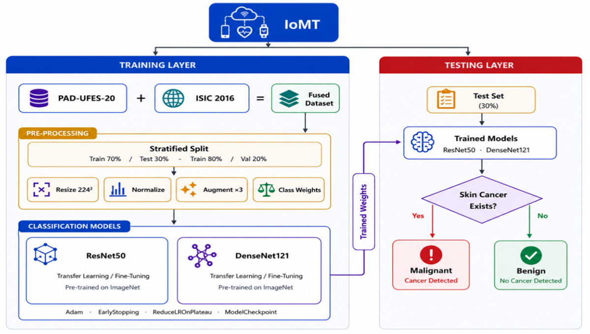
Skin cancer remains a major healthcare challenge. Early detection is important because it increases the chances of successful treatment. Deep learning has become an effective approach for automated skin lesion classification. However, many existing studies rely on a single dataset, which limits the diversity of training images and may reduce model generalization. This study proposes a transfer learning framework for binary skin cancer classification using dataset-level fusion. Two public datasets, PAD-UFES-20 and ISIC~2016, are combined to create a fused dataset that contains both clinical smartphone images and dermoscopic images. The images are resized to 224 × 224 pixels, converted to RGB format, and divided into training, validation, and testing sets using stratified splitting. Data augmentation and class weighting are applied to improve model learning. Two pretrained models, ResNet50 and DenseNet121, are fine-tuned using a two-phase training strategy. Model performance is evaluated using accuracy, sensitivity, specificity, positive and negative predictive values, AUC-ROC, and confusion matrices. Experimental results show that ResNet50 achieved an accuracy of 82.19%, while DenseNet121 achieved the best accuracy of 82.98%. The findings suggest that dataset-level fusion improves training data diversity and provides reliable performance for binary skin cancer classification. The proposed framework is organized into a Training Layer and a Testing Layer, which separate model development from inference. This design provides a flexible framework for future medical image classification applications.
Graphical Abstract
Keywords
Data Availability Statement
Funding
Conflicts of Interest
AI Use Statement
Ethical Approval and Consent to Participate
References
- Sung, H., Ferlay, J., Siegel, R. L., Laversanne, M., Soerjomataram, I., Jemal, A., & Bray, F. (2021). Global cancer statistics 2020: GLOBOCAN estimates of incidence and mortality worldwide for 36 cancers in 185 countries. CA: a cancer journal for clinicians, 71(3), 209-249.
[CrossRef] [Google Scholar] - Goyal, M., Knackstedt, T., Yan, S., & Hassanpour, S. (2020). Artificial intelligence-based image classification methods for diagnosis of skin cancer: Challenges and opportunities. Computers in biology and medicine, 127, 104065.
[CrossRef] [Google Scholar] - Fried, L., Tan, A., Bajaj, S., Liebman, T. N., Polsky, D., & Stein, J. A. (2020). Technological advances for the detection of melanoma: Advances in diagnostic techniques. Journal of the American Academy of Dermatology, 83(4), 983-992.
[CrossRef] [Google Scholar] - Janda, M., Cust, A. E., Neale, R. E., Aitken, J. F., Baade, P. D., Green, A. C., ... & Whiteman, D. C. (2020). Early detection of melanoma: a consensus report from the Australian skin and skin cancer research centre melanoma screening Summit. Australian and New Zealand journal of public health, 44(2), 111-115.
[CrossRef] [Google Scholar] - Davis, S., Piggott, C., Lyon, C., & DeSanto, K. (2020). Effectiveness of dermoscopy in skin cancer diagnosis. Canadian Family Physician, 66(10), 739-740. https://www.cfp.ca/content/66/10/739.short
[Google Scholar] - Sinz, C., Tschandl, P., Rosendahl, C., Akay, B. N., Argenziano, G., Blum, A., ... & Kittler, H. (2017). Accuracy of dermatoscopy for the diagnosis of nonpigmented cancers of the skin. Journal of the American Academy of Dermatology, 77(6), 1100-1109.
[CrossRef] [Google Scholar] - Healsmith, M. F., Bourke, J. F., Osborne, J. E., & Graham‐Brown, R. A. C. (1994). An evaluation of the revised seven‐point checklist for the early diagnosis of cutaneous malignant melanoma. British Journal of Dermatology, 130(1), 48-50.
[CrossRef] [Google Scholar] - Nachbar, F., Stolz, W., Merkle, T., Cognetta, A. B., Vogt, T., Landthaler, M., ... & Plewig, G. (1994). The ABCD rule of dermatoscopy: high prospective value in the diagnosis of doubtful melanocytic skin lesions. Journal of the American Academy of Dermatology, 30(4), 551-559.
[CrossRef] [Google Scholar] - Jung, J. M., Cho, J. Y., Lee, W. J., Chang, S. E., Lee, M. W., & Won, C. H. (2021). Emerging minimally invasive technologies for the detection of skin cancer. Journal of personalized medicine, 11(10), 951.
[CrossRef] [Google Scholar] - Esteva, A., Kuprel, B., Novoa, R. A., Ko, J., Swetter, S. M., Blau, H. M., & Thrun, S. (2017). Dermatologist-level classification of skin cancer with deep neural networks. nature, 542(7639), 115-118.
[CrossRef] [Google Scholar] - Haenssle, H. A., Fink, C., Schneiderbauer, R., Toberer, F., Buhl, T., Blum, A., ... & Zalaudek, I. (2018). Man against machine: diagnostic performance of a deep learning convolutional neural network for dermoscopic melanoma recognition in comparison to 58 dermatologists. Annals of oncology, 29(8), 1836-1842.
[CrossRef] [Google Scholar] - Kassem, M. A., Hosny, K. M., & Fouad, M. M. (2020). Skin lesions classification into eight classes for ISIC 2019 using deep convolutional neural network and transfer learning. IEEE Access, 8, 114822-114832.
[CrossRef] [Google Scholar] - Simonyan, K., & Zisserman, A. (2014). Very deep convolutional networks for large-scale image recognition. arXiv preprint arXiv:1409.1556.
[CrossRef] [Google Scholar] - Tan, M., & Le, Q. (2019, May). Efficientnet: Rethinking model scaling for convolutional neural networks. In International conference on machine learning (pp. 6105-6114). PmLR. https://proceedings.mlr.press/v97/tan19a.html?ref=ji
[Google Scholar] - Gutman, D., Codella, N. C., Celebi, E., Helba, B., Marchetti, M., Mishra, N., & Halpern, A. (2016). Skin lesion analysis toward melanoma detection: A challenge at the international symposium on biomedical imaging (ISBI) 2016, hosted by the international skin imaging collaboration (ISIC). arXiv preprint arXiv:1605.01397.
[CrossRef] [Google Scholar] - Pacheco, A. G., Lima, G. R., Salomao, A. S., Krohling, B., Biral, I. P., De Angelo, G. G., ... & De Barros, L. F. (2020). PAD-UFES-20: A skin lesion dataset composed of patient data and clinical images collected from smartphones. Data in brief, 32, 106221.
[CrossRef] [Google Scholar] - Tschandl, P., Rosendahl, C., & Kittler, H. (2018). The HAM10000 dataset, a large collection of multi-source dermatoscopic images of common pigmented skin lesions. Scientific data, 5(1), 180161.
[CrossRef] [Google Scholar] - Pacheco, A. G., & Krohling, R. A. (2021). An attention-based mechanism to combine images and metadata in deep learning models applied to skin cancer classification. IEEE journal of biomedical and health informatics, 25(9), 3554-3563.
[CrossRef] [Google Scholar] - Yin, W., Huang, J., Chen, J., & Ji, Y. (2022). A study on skin tumor classification based on dense convolutional networks with fused metadata. Frontiers in Oncology, 12, 989894.
[CrossRef] [Google Scholar] - Pacheco, A. G., & Krohling, R. A. (2020). The impact of patient clinical information on automated skin cancer detection. Computers in biology and medicine, 116, 103545.
[CrossRef] [Google Scholar] - Brinker, T. J., Hekler, A., Enk, A. H., Klode, J., Hauschild, A., Berking, C., ... & Schrüfer, P. (2019). A convolutional neural network trained with dermoscopic images performed on par with 145 dermatologists in a clinical melanoma image classification task. European journal of cancer, 111, 148-154.
[CrossRef] [Google Scholar] - de Lima, L. M., & Krohling, R. A. (2022, November). Exploring advances in transformers and CNN for skin lesion diagnosis on small datasets. In Brazilian Conference on Intelligent Systems (pp. 282-296). Cham: Springer International Publishing.
[CrossRef] [Google Scholar] - Tang, P., Yan, X., Nan, Y., Hu, X., Menze, B. H., Krammer, S., & Lasser, T. (2024). Joint-individual fusion structure with fusion attention module for multi-modal skin cancer classification. Pattern Recognition, 154, 110604.
[CrossRef] [Google Scholar] - Ge, Z., Demyanov, S., Chakravorty, R., Bowling, A., & Garnavi, R. (2017, September). Skin disease recognition using deep saliency features and multimodal learning of dermoscopy and clinical images. In International conference on medical image computing and computer-assisted intervention (pp. 250-258). Cham: Springer International Publishing.
[CrossRef] [Google Scholar] - Kawahara, J., Daneshvar, S., Argenziano, G., & Hamarneh, G. (2018). Seven-point checklist and skin lesion classification using multitask multimodal neural nets. IEEE journal of biomedical and health informatics, 23(2), 538-546.
[CrossRef] [Google Scholar] - Yap, J., Yolland, W., & Tschandl, P. (2018). Multimodal skin lesion classification using deep learning. Experimental dermatology, 27(11), 1261-1267.
[CrossRef] [Google Scholar] - Nunnari, F., Bhuvaneshwara, C., Ezema, A. O., & Sonntag, D. (2020, August). A study on the fusion of pixels and patient metadata in CNN-based classification of skin lesion images. In International cross-domain conference for machine learning and knowledge extraction (pp. 191-208). Cham: Springer International Publishing.
[CrossRef] [Google Scholar] - Chen, Q., Li, M., Chen, C., Zhou, P., Lv, X., & Chen, C. (2023). MDFNet: application of multimodal fusion method based on skin image and clinical data to skin cancer classification. Journal of Cancer Research and Clinical Oncology, 149(7), 3287-3299.
[CrossRef] [Google Scholar] - Ou, C., Zhou, S., Yang, R., Jiang, W., He, H., Gan, W., ... & Li, J. (2022). A deep learning based multimodal fusion model for skin lesion diagnosis using smartphone collected clinical images and metadata. Frontiers in Surgery, 9, 1029991.
[CrossRef] [Google Scholar] - Cai, G., Zhu, Y., Wu, Y., Jiang, X., Ye, J., & Yang, D. (2023). A multimodal transformer to fuse images and metadata for skin disease classification. The Visual Computer, 39(7), 2781-2793.
[CrossRef] [Google Scholar] - Tang, P., Yan, X., Nan, Y., Xiang, S., Krammer, S., & Lasser, T. (2022). FusionM4Net: A multi-stage multi-modal learning algorithm for multi-label skin lesion classification. Medical Image Analysis, 76, 102307.
[CrossRef] [Google Scholar] - Pedro, R., & Oliveira, A. L. (2022, July). Assessing the impact of attention and self-attention mechanisms on the classification of skin lesions. In 2022 international joint conference on neural networks (IJCNN) (pp. 1-8). IEEE.
[CrossRef] [Google Scholar] - Zhang, Y., Xie, F., & Chen, J. (2023). TFormer: A throughout fusion transformer for multi-modal skin lesion diagnosis. Computers in biology and medicine, 157, 106712.
[CrossRef] [Google Scholar] - Xu, J., Gao, Y., Liu, W., Huang, K., Zhao, S., Lu, L., ... & Chen, X. (2022, September). RemixFormer: a transformer model for precision skin tumor differential diagnosis via multi-modal imaging and non-imaging data. In International Conference on Medical Image Computing and Computer-Assisted Intervention (pp. 624-633). Cham: Springer Nature Switzerland.
[CrossRef] [Google Scholar] - Omeroglu, A. N., Mohammed, H. M., Oral, E. A., & Aydin, S. (2023). A novel soft attention-based multi-modal deep learning framework for multi-label skin lesion classification. Engineering Applications of Artificial Intelligence, 120, 105897.
[CrossRef] [Google Scholar] - Vachmanus, S., Noraset, T., Piyanonpong, W., Rattananukrom, T., & Tuarob, S. (2023). DeepMetaForge: A deep vision-transformer metadata-fusion network for automatic skin lesion classification. IEEE Access, 11, 145467-145484.
[CrossRef] [Google Scholar] - Cheslerean-Boghiu, T., Fleischmann, M. E., Willem, T., & Lasser, T. (2023). Transformer-based interpretable multi-modal data fusion for skin lesion classification. arXiv preprint arXiv:2304.14505.
[CrossRef] [Google Scholar] - Georgiadis, P., Gkouvrikos, E. V., Vrochidou, E., Kalampokas, T., & Papakostas, G. A. (2025). Building better deep learning models through dataset fusion: A case study in skin cancer classification with hyperdatasets. Diagnostics, 15(3), 352.
[CrossRef] [Google Scholar] - Remya, S., Anjali, T., & Sugumaran, V. (2024). A novel transfer learning framework for multimodal skin lesion analysis. IEEE Access, 12, 50738-50754.
[CrossRef] [Google Scholar] - Adebiyi, A., Abdalnabi, N., Simoes, E. J., Becevic, M., Hoffman Smith, E., & Rao, P. (2024). Transformers in skin lesion classification and diagnosis: A systematic review. MedRxiv, 2024-09.
[CrossRef] [Google Scholar] - Das, A., Agarwal, V., & Shetty, N. P. (2025). Comparative analysis of multimodal architectures for effective skin lesion detection using clinical and image data. Frontiers in Artificial Intelligence, 8, 1608837.
[CrossRef] [Google Scholar] - Marchetti, M. A., Codella, N. C., Dusza, S. W., Gutman, D. A., Helba, B., Kalloo, A., ... & International Skin Imaging Collaboration. (2018). Results of the 2016 International Skin Imaging Collaboration International Symposium on Biomedical Imaging challenge: Comparison of the accuracy of computer algorithms to dermatologists for the diagnosis of melanoma from dermoscopic images. Journal of the American Academy of Dermatology, 78(2), 270-277.
[CrossRef] [Google Scholar] - He, K., Zhang, X., Ren, S., & Sun, J. (2016). Deep residual learning for image recognition. In Proceedings of the IEEE conference on computer vision and pattern recognition (pp. 770-778).
[CrossRef] [Google Scholar] - Huang, G., Liu, Z., Van Der Maaten, L., & Weinberger, K. Q. (2017, July). Densely connected convolutional networks. In 2017 IEEE conference on computer vision and pattern recognition (CVPR) (pp. 2261-2269). IEEE.
[CrossRef] [Google Scholar] - Tajbakhsh, N., Shin, J. Y., Gurudu, S. R., Hurst, R. T., Kendall, C. B., Gotway, M. B., & Liang, J. (2016). Convolutional neural networks for medical image analysis: Full training or fine tuning?. IEEE transactions on medical imaging, 35(5), 1299-1312.
[CrossRef] [Google Scholar] - Uliana, J. J., & Krohling, R. A. (2025). Diffusion models applied to skin and oral cancer classification. arXiv preprint arXiv:2504.00026.
[CrossRef] [Google Scholar] - Azeem, M., Kiani, K., Mansouri, T., & Topping, N. (2023). SkinLesNet: classification of skin lesions and detection of melanoma cancer using a novel multi-layer deep convolutional neural network. Cancers, 16(1), 108.
[CrossRef] [Google Scholar] - Battle, M. L., Atapour-Abarghouei, A., & McGough, A. S. (2022, December). Siamese neural networks for skin cancer classification and new class detection using clinical and dermoscopic image datasets. In 2022 IEEE International Conference on Big Data (Big Data) (pp. 4346-4355). IEEE.
[CrossRef] [Google Scholar] - Khan, I. U., Aslam, N., Anwar, T., Aljameel, S. S., Ullah, M., Khan, R., ... & Akhtar, N. (2021). Remote diagnosis and triaging model for skin cancer using EfficientNet and extreme gradient boosting. Complexity, 2021(1), 5591614.
[CrossRef] [Google Scholar] - Saxena, S., Kaushik, K., & Chaudhary, A. (2025). AI-Driven Early Detection of Skin Cancer using PAD-UFES-20: TRL 1 & 2 Achievements in Multimodal Deep Learning.
[CrossRef] [Google Scholar]
Cite This Article
TY  - JOUR
AU  - Mubarik, Tuba
AU  - Aftab, Shabib
PY  - 2026
DA  - 2026/10/09
TI  - Skin Cancer Detection Using Data Fusion and Deep Transfer Learning Techniques
JO  - ICCK Journal of Image Analysis and Processing
T2  - ICCK Journal of Image Analysis and Processing
JF  - ICCK Journal of Image Analysis and Processing
VL  - 2
IS  - 4
SP  - 218
EP  - 230
DO  - 10.62762/JIAP.2026.243543
UR  - https://www.icck.org/article/abs/JIAP.2026.243543
KW  - Skin cancer
KW  - binary classification
KW  - dataset fusion
KW  - transfer learning
KW  - ResNet50
KW  - DenseNet121
KW  - PAD-UFES-20
KW  - ISIC 2016
AB  - Skin cancer remains a major healthcare challenge. Early detection is important because it increases the chances of successful treatment. Deep learning has become an effective approach for automated skin lesion classification. However, many existing studies rely on a single dataset, which limits the diversity of training images and may reduce model generalization. This study proposes a transfer learning framework for binary skin cancer classification using dataset-level fusion. Two public datasets, PAD-UFES-20 and ISIC~2016, are combined to create a fused dataset that contains both clinical smartphone images and dermoscopic images. The images are resized to 224 × 224 pixels, converted to RGB format, and divided into training, validation, and testing sets using stratified splitting. Data augmentation and class weighting are applied to improve model learning. Two pretrained models, ResNet50 and DenseNet121, are fine-tuned using a two-phase training strategy. Model performance is evaluated using accuracy, sensitivity, specificity, positive and negative predictive values, AUC-ROC, and confusion matrices. Experimental results show that ResNet50 achieved an accuracy of 82.19%, while DenseNet121 achieved the best accuracy of 82.98%. The findings suggest that dataset-level fusion improves training data diversity and provides reliable performance for binary skin cancer classification. The proposed framework is organized into a Training Layer and a Testing Layer, which separate model development from inference. This design provides a flexible framework for future medical image classification applications.
SN  - 3068-6679
PB  - Institute of Central Computation and Knowledge
LA  - English
ER  -
@article{Mubarik2026Skin,
  author = {Tuba Mubarik and Shabib Aftab},
  title = {Skin Cancer Detection Using Data Fusion and Deep Transfer Learning Techniques},
  journal = {ICCK Journal of Image Analysis and Processing},
  year = {2026},
  volume = {2},
  number = {4},
  pages = {218-230},
  doi = {10.62762/JIAP.2026.243543},
  url = {https://www.icck.org/article/abs/JIAP.2026.243543},
  abstract = {Skin cancer remains a major healthcare challenge. Early detection is important because it increases the chances of successful treatment. Deep learning has become an effective approach for automated skin lesion classification. However, many existing studies rely on a single dataset, which limits the diversity of training images and may reduce model generalization. This study proposes a transfer learning framework for binary skin cancer classification using dataset-level fusion. Two public datasets, PAD-UFES-20 and ISIC~2016, are combined to create a fused dataset that contains both clinical smartphone images and dermoscopic images. The images are resized to 224 × 224 pixels, converted to RGB format, and divided into training, validation, and testing sets using stratified splitting. Data augmentation and class weighting are applied to improve model learning. Two pretrained models, ResNet50 and DenseNet121, are fine-tuned using a two-phase training strategy. Model performance is evaluated using accuracy, sensitivity, specificity, positive and negative predictive values, AUC-ROC, and confusion matrices. Experimental results show that ResNet50 achieved an accuracy of 82.19\%, while DenseNet121 achieved the best accuracy of 82.98\%. The findings suggest that dataset-level fusion improves training data diversity and provides reliable performance for binary skin cancer classification. The proposed framework is organized into a Training Layer and a Testing Layer, which separate model development from inference. This design provides a flexible framework for future medical image classification applications.},
  keywords = {Skin cancer, binary classification, dataset fusion, transfer learning, ResNet50, DenseNet121, PAD-UFES-20, ISIC 2016},
  issn = {3068-6679},
  publisher = {Institute of Central Computation and Knowledge}
}
Article Metrics
Publisher's Note
ICCK stays neutral with regard to jurisdictional claims in published maps and institutional affiliations.
Rights and Permissions
 Copyright © 2026 by the Author(s). Published by Institute of Central Computation and Knowledge. This article is an open access article distributed under the terms and conditions of the Creative Commons Attribution (CC BY) license (https://creativecommons.org/licenses/by/4.0/), which permits use, sharing, adaptation, distribution and reproduction in any medium or format, as long as you give appropriate credit to the original author(s) and the source, provide a link to the Creative Commons licence, and indicate if changes were made.


Portico
