CT Image Denoising using Discrete Wavelet Transform

Article Information
Abstract
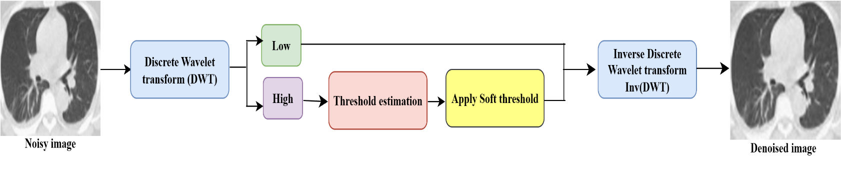
Low Dose Computed Tomography (LDCT) scan is modern medical imaging diagnostic technique that provides a detailed projection of internal human body tissue level structures. Even though the LDCT image quality is compromised by Gaussian-noise, which can be generated during image acquisition, this compromises the accurate diagnostic precision. The effective denoising is required to improve image quality in LDCT images. This study demonstrates that the Discrete Wavelet Transform(DWT) method shows better results, both quantitatively and visually, under varying noise intensities ($\sigma=10,20,30,$ and $40$). The DWT method decomposes the image to multiresolution subbands (approximation, and detail) to provide localized analysis of structural patterns. The thresholding method is applied to the detail (noisy) coefficients and then reconstructs the refined image from these denoised coefficients. The DWT method achieved superior noise suppression while preserving edge information. The quantitative analysis among various methods, including PCA, MSVD, DCT, and DWT, consistently shows superior results, achieving a higher PSNR of $33.85$ dB, SNR of $28.50$ dB, and SSIM of $0.7194$ at a noise level $\sigma =10$. Among all denoising methods, the DWT is a powerful and consistent method in image processing to enhance image quality in LDCT images.
Graphical Abstract
Keywords
Data Availability Statement
Funding
Conflicts of Interest
Ethical Approval and Consent to Participate
References
- Sadia, R. T., Chen, J., & Zhang, J. (2024). CT image denoising methods for image quality improvement and radiation dose reduction. Journal of applied clinical medical physics, 25(2), e14270.
[CrossRef] [Google Scholar] - Abuya, T. K., Rimiru, R. M., & Okeyo, G. O. (2023). An image denoising technique using wavelet-anisotropic gaussian filter-based denoising convolutional neural network for CT images. Applied sciences, 13(21), 12069.
[CrossRef] [Google Scholar] - Kumar, R. R., & Priyadarshi, R. (2025). Denoising and segmentation in medical image analysis: A comprehensive review on machine learning and deep learning approaches. Multimedia Tools and Applications, 84(12), 10817-10875.
[CrossRef] [Google Scholar] - Zhang, F., Liu, J., Liu, Y., & Zhang, X. (2023). Research progress of deep learning in low-dose CT image denoising. Radiation protection dosimetry, 199(4), 337-346.
[CrossRef] [Google Scholar] - Mao, J., Sun, L., Chen, J., & Yu, S. (2025). Overview of Research on Digital Image Denoising Methods. Sensors, 25(8), 2615.
[CrossRef] [Google Scholar] - Zhou, Y., Kong, Z., Huang, T., Ahn, E., Li, H., & Ding, L. (2024). WaveletDFDS-Net: A Dual Forward Denoising Stream Network for Low-Dose CT Noise Reduction. Electronics, 13(10), 1906.
[CrossRef] [Google Scholar] - Esfahani, E. E., & Gouran, A. (2025). Low-dose CT using a nonlocal and nonlinear principal component analysis for image restoration. IEEE Transactions on Radiation and Plasma Medical Sciences.
[CrossRef] [Google Scholar] - Hosen, M. A., Moz, S. H., Kabir, S. S., Adnan, M. N., & Galib, S. M. (2024). In-depth exploration of digital image watermarking with discrete cosine transform and discrete wavelet transform. Indonesian Journal of Electrical Engineering and Computer Science, 33(1), 581-90.
[CrossRef] [Google Scholar] - Katageri, G. S., & Swamy, P. S. (2025). Denoising and analysis of synthetic aperture radar images using improved weight threshold technique in curvelet transform frequency domain. Multimedia Tools and Applications, 84(12), 10173-10194.
[CrossRef] [Google Scholar] - Bhosekar, S., Singh, P., & Garg, D. (2025, January). A Comparative Analysis of Multi-Modal Medical Image Fusion Techniques using MSVD, WPD, PCA, and DWT. In 2025 International Conference on Cognitive Computing in Engineering, Communications, Sciences and Biomedical Health Informatics (IC3ECSBHI) (pp. 923-927). IEEE.
[CrossRef] [Google Scholar] - Alnuaimy, A. N., Jawad, A. M., Abdulkareem, S. A., Mustafa, F. M., Ivanchenko, S., & Toliupa, S. (2024, April). Bm3d denoising algorithms for medical image. In 2024 35th Conference of Open Innovations Association (FRUCT) (pp. 135-141). IEEE.
[CrossRef] [Google Scholar] - Choi, K. (2024). Self-supervised learning for CT image denoising and reconstruction: a review. Biomedical Engineering Letters, 14(6), 1207-1220.
[CrossRef] [Google Scholar] - Lei, Y., Niu, C., Zhang, J., Wang, G., & Shan, H. (2023). CT image denoising and deblurring with deep learning: current status and perspectives. IEEE Transactions on Radiation and Plasma Medical Sciences, 8(2), 153-172.
[CrossRef] [Google Scholar] - Zhang, B., Zhang, Y., Wang, B., He, X., Zhang, F., & Zhang, X. (2024). Denoising swin transformer and perceptual peak signal-to-noise ratio for low-dose CT image denoising. Measurement, 227, 114303.
[CrossRef] [Google Scholar] - Yuan, J., Zhou, F., Guo, Z., Li, X., & Yu, H. (2023). HCformer: hybrid CNN-transformer for LDCT image denoising. Journal of Digital Imaging, 36(5), 2290-2305.
[CrossRef] [Google Scholar] - Soares, E., Angelov, P., Biaso, S., Froes, M. H., & Abe, D. K. (2020). SARS-CoV-2 CT-scan dataset: A large dataset of real patients CT scans for SARS-CoV-2 identification. MedRxiv, 2020-04.
[CrossRef] [Google Scholar] - Zubair, M., Helmi, B., Ullah, F., Al-Tashi, Q., Faheem, M., & Khan, A. A. (2024). Enabling predication of the deep learning algorithms for low-dose CT scan image denoising models: A systematic literature review. IEEE Access, 12, 79025-79050.
[CrossRef] [Google Scholar]
Cited By (1)
-
Sai Bhargav Kasetty, Rajakumar Krishnan. A unified comparative framework for multiscale geometric transforms in SAR and multispectral satellite image analysis.
Frontiers in Remote Sensing, 2026 , 7 .
[CrossRef]
Cite This Article
TY  - JOUR
AU  - Katta, Swapna
AU  - Garg, Deepak
PY  - 2025
DA  - 2025/09/22
TI  - CT Image Denoising using Discrete Wavelet Transform
JO  - Biomedical Informatics and Smart Healthcare
T2  - Biomedical Informatics and Smart Healthcare
JF  - Biomedical Informatics and Smart Healthcare
VL  - 1
IS  - 2
SP  - 44
EP  - 51
DO  - 10.62762/BISH.2025.874472
UR  - https://www.icck.org/article/abs/BISH.2025.874472
KW  - CT image
KW  - denoising Gaussian noise
KW  - DWT
KW  - Transform domain
AB  - Low Dose Computed Tomography (LDCT) scan is modern medical imaging diagnostic technique that provides a detailed projection of internal human body tissue level structures. Even though the LDCT image quality is compromised by Gaussian-noise, which can be generated during image acquisition, this compromises the accurate diagnostic precision. The effective denoising is required to improve image quality in LDCT images. This study demonstrates that the Discrete Wavelet Transform(DWT) method shows better results, both quantitatively and visually, under varying noise intensities ($\sigma=10,20,30,$ and $40$). The DWT method decomposes the image to multiresolution subbands (approximation, and detail) to provide localized analysis of structural patterns. The thresholding method is applied to the detail (noisy) coefficients and then reconstructs the refined image from these denoised coefficients. The DWT method achieved superior noise suppression while preserving edge information. The quantitative analysis among various methods, including PCA, MSVD, DCT, and DWT, consistently shows superior results, achieving a higher PSNR of $33.85$ dB, SNR of $28.50$ dB, and SSIM of $0.7194$ at a noise level $\sigma =10$. Among all denoising methods, the DWT is a powerful and consistent method in image processing to enhance image quality in LDCT images.
SN  - 3068-5524
PB  - Institute of Central Computation and Knowledge
LA  - English
ER  -
@article{Katta2025CT,
  author = {Swapna Katta and Deepak Garg},
  title = {CT Image Denoising using Discrete Wavelet Transform},
  journal = {Biomedical Informatics and Smart Healthcare},
  year = {2025},
  volume = {1},
  number = {2},
  pages = {44-51},
  doi = {10.62762/BISH.2025.874472},
  url = {https://www.icck.org/article/abs/BISH.2025.874472},
  abstract = {Low Dose Computed Tomography (LDCT) scan is modern medical imaging diagnostic technique that provides a detailed projection of internal human body tissue level structures. Even though the LDCT image quality is compromised by Gaussian-noise, which can be generated during image acquisition, this compromises the accurate diagnostic precision. The effective denoising is required to improve image quality in LDCT images. This study demonstrates that the Discrete Wavelet Transform(DWT) method shows better results, both quantitatively and visually, under varying noise intensities (\$\sigma=10,20,30,\$ and \$40\$). The DWT method decomposes the image to multiresolution subbands (approximation, and detail) to provide localized analysis of structural patterns. The thresholding method is applied to the detail (noisy) coefficients and then reconstructs the refined image from these denoised coefficients. The DWT method achieved superior noise suppression while preserving edge information. The quantitative analysis among various methods, including PCA, MSVD, DCT, and DWT, consistently shows superior results, achieving a higher PSNR of \$33.85\$ dB, SNR of \$28.50\$ dB, and SSIM of \$0.7194\$ at a noise level \$\sigma =10\$. Among all denoising methods, the DWT is a powerful and consistent method in image processing to enhance image quality in LDCT images.},
  keywords = {CT image, denoising Gaussian noise, DWT, Transform domain},
  issn = {3068-5524},
  publisher = {Institute of Central Computation and Knowledge}
}
Publisher's Note
ICCK stays neutral with regard to jurisdictional claims in published maps and institutional affiliations.
Rights and Permissions
 Copyright © 2025 by the Author(s). Published by Institute of Central Computation and Knowledge. This article is an open access article distributed under the terms and conditions of the Creative Commons Attribution (CC BY) license (https://creativecommons.org/licenses/by/4.0/), which permits use, sharing, adaptation, distribution and reproduction in any medium or format, as long as you give appropriate credit to the original author(s) and the source, provide a link to the Creative Commons licence, and indicate if changes were made.

Portico
