Efficient Polyp Segmentation via Attention-Guided Lightweight Network with Progressive Multi-Scale Fusion
Research Article  ·  Published: 05 June 2025
Issue cover
ICCK Transactions on Intelligent Systematics
Volume 2, Issue 2, 2025: 95-108
Research Article Free to Read

Efficient Polyp Segmentation via Attention-Guided Lightweight Network with Progressive Multi-Scale Fusion

1 Department of Pharmacy, University of Bradford, Bradford, BD7 1DP, United Kingdom
2 Health Services Management Department, University of Chester, Chester CH1 4BJ, United Kingdom
3 Mardan Medical Complex, Mardan 23200, Pakistan
4 Northwest School of Medicine, Peshawar 25000, Pakistan
5 Departamento de Sistemas Informaticos, Universidad Politécnica de Madrid, Madrid 28031, Spain
* Corresponding Author: Muhammad Jamal Ahmed, [email protected]
Volume 2, Issue 2
You have access to this article · Limited-Time Free Access

Article Information

Abstract

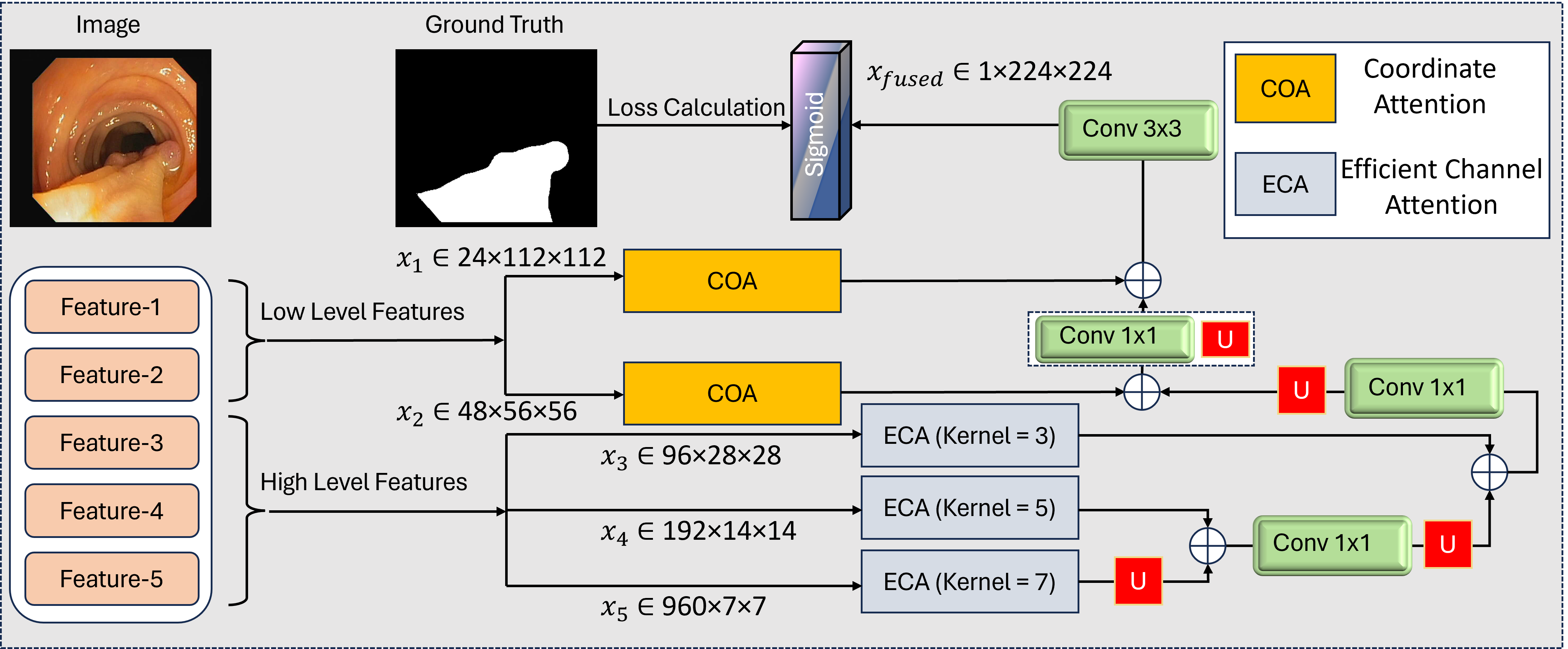
Accurate and real-time polyp segmentation plays a vital role in the early detection of colorectal cancer. However, existing methods often rely on computationally expensive backbones, single attention mechanisms, and suboptimal feature fusion strategies, limiting their practicality in real-world scenarios. In this work, we propose a lightweight yet effective deep learning framework that strikes a balance between precision and efficiency through a carefully designed architecture. Specifically, we adopt a MobileNetV4-based hybrid backbone to extract rich multi-scale features with significantly fewer parameters than conventional backbones, making the model well-suited for resource-constrained clinical settings. To enhance feature representation, we introduce a novel dual-attention guidance mechanism that integrates Efficient Channel Attention (ECA) for channel-wise refinement and Coordinate Attention (COA) for spatial modeling, which is particularly effective at delineating polyp boundaries. Additionally, we design a progressive multi-scale fusion strategy that hierarchically integrates feature maps from deep to shallow layers, preserving spatial details while enhancing contextual understanding. Extensive experiments on five benchmark polyp segmentation datasets demonstrate that our method consistently outperforms state-of-the-art approaches across both quantitative metrics and qualitative visualizations. Comprehensive ablation studies further validate the effectiveness of each component, highlighting the practical viability of our approach for real-time polyp segmentation applications.

Graphical Abstract

Efficient Polyp Segmentation via Attention-Guided Lightweight Network with Progressive Multi-Scale Fusion

Keywords

colorectal cancer visual intelligence polyp segmentation lightweight network dual attention multi-scale fusion medical imaging

Data Availability Statement

Data will be made available on request.

Funding

This work was supported without any funding.

Conflicts of Interest

The authors declare no conflicts of interest.

Ethical Approval and Consent to Participate

This study utilized only publicly available, anonymized datasets. Therefore, ethical approval was not required according to institutional and national guidelines.

References

  1. Hossain, M. S., Karuniawati, H., Jairoun, A. A., Urbi, Z., Ooi, D. J., John, A., ... & Hadi, M. A. (2022). Colorectal cancer: a review of carcinogenesis, global epidemiology, current challenges, risk factors, preventive and treatment strategies. Cancers, 14(7), 1732.
    [CrossRef] [Google Scholar]
  2. Kim, N. H., Jung, Y. S., Jeong, W. S., Yang, H. J., Park, S. K., Choi, K., & Park, D. I. (2017). Miss rate of colorectal neoplastic polyps and risk factors for missed polyps in consecutive colonoscopies. Intestinal research, 15(3), 411.
    [Google Scholar]
  3. Prasath, V. S. (2016). Polyp detection and segmentation from video capsule endoscopy: A review. Journal of Imaging, 3(1), 1.
    [CrossRef] [Google Scholar]
  4. Zhao, X., Jia, H., Pang, Y., Lv, L., Tian, F., Zhang, L., ... & Lu, H. (2). M2SNet: Multi-scale in Multi-scale Subtraction Network for Medical Image Segmentation. arXiv 2023. arXiv preprint arXiv:2303.10894.
    [Google Scholar]
  5. Patel, K., Bur, A. M., & Wang, G. (2021, May). Enhanced u-net: A feature enhancement network for polyp segmentation. In 2021 18th conference on robots and vision (CRV) (pp. 181-188). IEEE.
    [CrossRef] [Google Scholar]
  6. Zhou, T., Zhou, Y., He, K., Gong, C., Yang, J., Fu, H., & Shen, D. (2023). Cross-level feature aggregation network for polyp segmentation. Pattern Recognition, 140, 109555.
    [CrossRef] [Google Scholar]
  7. Yue, G., Han, W., Jiang, B., Zhou, T., Cong, R., & Wang, T. (2022). Boundary constraint network with cross layer feature integration for polyp segmentation. IEEE Journal of Biomedical and Health Informatics, 26(8), 4090-4099.
    [CrossRef] [Google Scholar]
  8. Yang, H., Chen, Q., Fu, K., Zhu, L., Jin, L., Qiu, B., ... & Lu, Y. (2022). Boosting medical image segmentation via conditional-synergistic convolution and lesion decoupling. Computerized Medical Imaging and Graphics, 101, 102110.
    [CrossRef] [Google Scholar]
  9. Long, J., Shelhamer, E., & Darrell, T. (2015). Fully convolutional networks for semantic segmentation. In Proceedings of the IEEE conference on computer vision and pattern recognition (pp. 3431-3440).
    [Google Scholar]
  10. Ronneberger, O., Fischer, P., & Brox, T. (2015). U-net: Convolutional networks for biomedical image segmentation. In Medical image computing and computer-assisted intervention–MICCAI 2015: 18th international conference, Munich, Germany, October 5-9, 2015, proceedings, part III 18 (pp. 234-241). Springer international publishing.
    [CrossRef] [Google Scholar]
  11. Zhou, Z., Rahman Siddiquee, M. M., Tajbakhsh, N., & Liang, J. (2018). Unet++: A nested u-net architecture for medical image segmentation. In Deep learning in medical image analysis and multimodal learning for clinical decision support: 4th international workshop, DLMIA 2018, and 8th international workshop, ML-CDS 2018, held in conjunction with MICCAI 2018, Granada, Spain, September 20, 2018, proceedings 4 (pp. 3-11). Springer International Publishing.
    [CrossRef] [Google Scholar]
  12. Fang, Y., Chen, C., Yuan, Y., & Tong, K. Y. (2019). Selective feature aggregation network with area-boundary constraints for polyp segmentation. In Medical Image Computing and Computer Assisted Intervention–MICCAI 2019: 22nd International Conference, Shenzhen, China, October 13–17, 2019, Proceedings, Part I 22 (pp. 302-310). Springer International Publishing.
    [CrossRef] [Google Scholar]
  13. Hatamizadeh, A., Terzopoulos, D., & Myronenko, A. (2019, October). End-to-end boundary aware networks for medical image segmentation. In International Workshop on Machine Learning in Medical Imaging (pp. 187-194). Cham: Springer International Publishing.
    [CrossRef] [Google Scholar]
  14. Chen, J., Lu, Y., Yu, Q., Luo, X., Adeli, E., Wang, Y., ... & Zhou, Y. (2021). Transunet: Transformers make strong encoders for medical image segmentation. arXiv preprint arXiv:2102.04306.
    [Google Scholar]
  15. Valanarasu, J. M. J., Oza, P., Hacihaliloglu, I., & Patel, V. M. (2021). Medical transformer: Gated axial-attention for medical image segmentation. In Medical image computing and computer assisted intervention–MICCAI 2021: 24th international conference, Strasbourg, France, September 27–October 1, 2021, proceedings, part I 24 (pp. 36-46). Springer International Publishing.
    [CrossRef] [Google Scholar]
  16. Wang, Y., Zhou, Q., Liu, J., Xiong, J., Gao, G., Wu, X., & Latecki, L. J. (2019, September). Lednet: A lightweight encoder-decoder network for real-time semantic segmentation. In 2019 IEEE international conference on image processing (ICIP) (pp. 1860-1864). IEEE.
    [CrossRef] [Google Scholar]
  17. Zhao, X., Zhang, L., & Lu, H. (2021). Automatic polyp segmentation via multi-scale subtraction network. In Medical Image Computing and Computer Assisted Intervention–MICCAI 2021: 24th International Conference, Strasbourg, France, September 27–October 1, 2021, Proceedings, Part I 24 (pp. 120-130). Springer International Publishing.
    [CrossRef] [Google Scholar]
  18. Fan, D. P., Ji, G. P., Zhou, T., Chen, G., Fu, H., Shen, J., & Shao, L. (2020, September). Pranet: Parallel reverse attention network for polyp segmentation. In International conference on medical image computing and computer-assisted intervention (pp. 263-273). Cham: Springer International Publishing.
    [CrossRef] [Google Scholar]
  19. Jha, D., Smedsrud, P. H., Riegler, M. A., Johansen, D., De Lange, T., Halvorsen, P., & Johansen, H. D. (2019, December). Resunet++: An advanced architecture for medical image segmentation. In 2019 IEEE international symposium on multimedia (ISM) (pp. 225-2255). IEEE.
    [CrossRef] [Google Scholar]
  20. Chen, W., Wang, K., Qian, C., Li, X., Li, C., & Duan, X. (2024). PPFormer: A novel model for polyp segmentation in digestive endoscopy. IEEE Transactions on Medical Robotics and Bionics, 6(2), 548-555.
    [CrossRef] [Google Scholar]
  21. Dong, B., Wang, W., Fan, D. P., Li, J., Fu, H., & Shao, L. (2021). Polyp-pvt: Polyp segmentation with pyramid vision transformers. arXiv preprint arXiv:2108.06932.
    [Google Scholar]
  22. Wei, J., Hu, Y., Zhang, R., Li, Z., Zhou, S. K., & Cui, S. (2021, September). Shallow attention network for polyp segmentation. In International conference on medical image computing and computer-assisted intervention (pp. 699-708). Cham: Springer International Publishing.
    [CrossRef] [Google Scholar]
  23. Murugesan, B., Sarveswaran, K., Shankaranarayana, S. M., Ram, K., Joseph, J., & Sivaprakasam, M. (2019, July). Psi-Net: Shape and boundary aware joint multi-task deep network for medical image segmentation. In 2019 41st Annual international conference of the IEEE engineering in medicine and biology society (EMBC) (pp. 7223-7226). IEEE.
    [CrossRef] [Google Scholar]
  24. Li, R., Su, J., Duan, C., & Zheng, S. (2020). Linear attention mechanism: An efficient attention for semantic segmentation. arXiv preprint arXiv:2007.14902.
    [Google Scholar]
  25. Nguyen, Q. V., Vo, T. H. S., Kang, S. R., & Kim, S. H. (2024). Polyp-SES: Automatic Polyp Segmentation with Self-Enriched Semantic Model. In Proceedings of the Asian Conference on Computer Vision (pp. 2803-2819).
    [Google Scholar]
  26. Wu, H., Zhao, Z., Zhong, J., Wang, W., Wen, Z., & Qin, J. (2022). Polypseg+: A lightweight context-aware network for real-time polyp segmentation. IEEE Transactions on Cybernetics, 53(4), 2610-2621.
    [CrossRef] [Google Scholar]
  27. Ji, Z., Qian, H., & Ma, X. (2024). Progressive group convolution fusion network for colon polyp segmentation. Biomedical Signal Processing and Control, 96, 106586.
    [CrossRef] [Google Scholar]
  28. Fang, X., & Yan, P. (2020). Multi-organ segmentation over partially labeled datasets with multi-scale feature abstraction. IEEE Transactions on Medical Imaging, 39(11), 3619-3629.
    [CrossRef] [Google Scholar]
  29. He, J., Deng, Z., & Qiao, Y. (2019). Dynamic multi-scale filters for semantic segmentation. In Proceedings of the IEEE/CVF international conference on computer vision (pp. 3562-3572).
    [Google Scholar]
  30. Sinha, A., & Dolz, J. (2020). Multi-scale self-guided attention for medical image segmentation. IEEE journal of biomedical and health informatics, 25(1), 121-130.
    [CrossRef] [Google Scholar]
  31. Hou, Q., Zhou, D., & Feng, J. (2021). Coordinate attention for efficient mobile network design. In Proceedings of the IEEE/CVF conference on computer vision and pattern recognition (pp. 13713-13722).
    [Google Scholar]
  32. Hu, J., Shen, L., & Sun, G. (2018). Squeeze-and-excitation networks. In Proceedings of the IEEE conference on computer vision and pattern recognition (pp. 7132-7141).
    [Google Scholar]
  33. Jha, D., Smedsrud, P. H., Riegler, M. A., Halvorsen, P., De Lange, T., Johansen, D., & Johansen, H. D. (2019, December). Kvasir-seg: A segmented polyp dataset. In International conference on multimedia modeling (pp. 451-462). Cham: Springer International Publishing.
    [CrossRef] [Google Scholar]
  34. Bernal, J., Sánchez, F. J., Fernández-Esparrach, G., Gil, D., Rodríguez, C., & Vilariño, F. (2015). WM-DOVA maps for accurate polyp highlighting in colonoscopy: Validation vs. saliency maps from physicians. Computerized medical imaging and graphics, 43, 99-111.
    [CrossRef] [Google Scholar]
  35. Tajbakhsh, N., Gurudu, S. R., & Liang, J. (2015). Automated polyp detection in colonoscopy videos using shape and context information. IEEE transactions on medical imaging, 35(2), 630-644.
    [CrossRef] [Google Scholar]
  36. Vázquez, D., Bernal, J., Sánchez, F. J., Fernández-Esparrach, G., López, A. M., Romero, A., ... & Courville, A. (2017). A benchmark for endoluminal scene segmentation of colonoscopy images. Journal of healthcare engineering, 2017(1), 4037190.
    [CrossRef] [Google Scholar]
  37. Silva, J., Histace, A., Romain, O., Dray, X., & Granado, B. (2014). Toward embedded detection of polyps in wce images for early diagnosis of colorectal cancer. International journal of computer assisted radiology and surgery, 9, 283-293.
    [CrossRef] [Google Scholar]
  38. Zhang, R., Li, G., Li, Z., Cui, S., Qian, D., & Yu, Y. (2020). Adaptive context selection for polyp segmentation. In Medical Image Computing and Computer Assisted Intervention–MICCAI 2020: 23rd International Conference, Lima, Peru, October 4–8, 2020, Proceedings, Part VI 23 (pp. 253-262). Springer International Publishing.
    [CrossRef] [Google Scholar]
  39. Kim, T., Lee, H., & Kim, D. (2021, October). Uacanet: Uncertainty augmented context attention for polyp segmentation. In Proceedings of the 29th ACM international conference on multimedia (pp. 2167-2175).
    [CrossRef] [Google Scholar]
  40. Qiu, Z., Wang, Z., Zhang, M., Xu, Z., Fan, J., & Xu, L. (2022, April). BDG-Net: boundary distribution guided network for accurate polyp segmentation. In Medical Imaging 2022: Image Processing (Vol. 12032, pp. 792-799). SPIE.
    [CrossRef] [Google Scholar]
  41. Wang, J., Huang, Q., Tang, F., Meng, J., Su, J., & Song, S. (2022, September). Stepwise feature fusion: Local guides global. In International conference on medical image computing and computer-assisted intervention (pp. 110-120). Cham: Springer Nature Switzerland.
    [CrossRef] [Google Scholar]
  42. Bui, N. T., Hoang, D. H., Nguyen, Q. T., Tran, M. T., & Le, N. (2024). Meganet: Multi-scale edge-guided attention network for weak boundary polyp segmentation. In Proceedings of the IEEE/CVF Winter Conference on Applications of Computer Vision (pp. 7985-7994).
    [Google Scholar]

Cited By (4)

  1. Bin Sun, Tao Hu, Fuhua Zhang, Leyuan Fang, Shutao Li. Multi-to-Uni modality knowledge distillation for remote sensing image semantic segmentation. Information Fusion, 2027 , 138 .
    [CrossRef]
  2. Peng Shi, Jingjing Guo, Lu Deng, Yingkai Liu, Lizhi Long, Shaopeng Xu. Enhanced recognition of low-discernibility Railway Sleeper Serial Numbers via dual-stage adaptive image enhancement and position prior-guided detection. Engineering Applications of Artificial Intelligence, 2026 , 169 .
    [CrossRef]
  3. Zhiwei Feng, Benyi Yang, Baosong Deng. SAM-assisted multimodal collaborative enhancement for remote sensing image segmentation. Information Fusion, 2026 , 135 .
    [CrossRef]
  4. Chengpeng Tian, Yuanyuan Peng, Yan Xi, Bin Tang, Qingkai Ma, Na Cheng, Zhen Liang, Kai Xu. A parallel hybrid convolutional state space network with multi-attention mechanisms for retinal vessel segmentation. Biomedical Signal Processing and Control, 2026 , 116 .
    [CrossRef]
* Citation data provided by Crossref Cited-by.

Cite This Article

APA Style
Mohammed, E., Khan, A., Ullah, W., Khan, W., & Ahmed, M. J. (2025). Efficient Polyp Segmentation via Attention-Guided Lightweight Network with Progressive Multi-Scale Fusion. ICCK Transactions on Intelligent Systematics, 2(2), 95-108. https://doi.org/10.62762/TIS.2025.389995
Export Citation
RIS Format
Compatible with EndNote, Zotero, Mendeley, and other reference managers
TY  - JOUR
AU  - Mohammed, Essa
AU  - Khan, Abdullah
AU  - Ullah, Waqas
AU  - Khan, Wisal
AU  - Ahmed, Muhammad Jamal
PY  - 2025
DA  - 2025/06/05
TI  - Efficient Polyp Segmentation via Attention-Guided Lightweight Network with Progressive Multi-Scale Fusion
JO  - ICCK Transactions on Intelligent Systematics
T2  - ICCK Transactions on Intelligent Systematics
JF  - ICCK Transactions on Intelligent Systematics
VL  - 2
IS  - 2
SP  - 95
EP  - 108
DO  - 10.62762/TIS.2025.389995
UR  - https://www.icck.org/article/abs/TIS.2025.389995
KW  - colorectal cancer
KW  - visual intelligence
KW  - polyp segmentation
KW  - lightweight network
KW  - dual attention
KW  - multi-scale fusion
KW  - medical imaging
AB  - Accurate and real-time polyp segmentation plays a vital role in the early detection of colorectal cancer. However, existing methods often rely on computationally expensive backbones, single attention mechanisms, and suboptimal feature fusion strategies, limiting their practicality in real-world scenarios. In this work, we propose a lightweight yet effective deep learning framework that strikes a balance between precision and efficiency through a carefully designed architecture. Specifically, we adopt a MobileNetV4-based hybrid backbone to extract rich multi-scale features with significantly fewer parameters than conventional backbones, making the model well-suited for resource-constrained clinical settings. To enhance feature representation, we introduce a novel dual-attention guidance mechanism that integrates Efficient Channel Attention (ECA) for channel-wise refinement and Coordinate Attention (COA) for spatial modeling, which is particularly effective at delineating polyp boundaries. Additionally, we design a progressive multi-scale fusion strategy that hierarchically integrates feature maps from deep to shallow layers, preserving spatial details while enhancing contextual understanding. Extensive experiments on five benchmark polyp segmentation datasets demonstrate that our method consistently outperforms state-of-the-art approaches across both quantitative metrics and qualitative visualizations. Comprehensive ablation studies further validate the effectiveness of each component, highlighting the practical viability of our approach for real-time polyp segmentation applications.
SN  - 3068-5079
PB  - Institute of Central Computation and Knowledge
LA  - English
ER  - 
BibTeX Format
Compatible with LaTeX, BibTeX, and other reference managers
@article{Mohammed2025Efficient,
  author = {Essa Mohammed and Abdullah Khan and Waqas Ullah and Wisal Khan and Muhammad Jamal Ahmed},
  title = {Efficient Polyp Segmentation via Attention-Guided Lightweight Network with Progressive Multi-Scale Fusion},
  journal = {ICCK Transactions on Intelligent Systematics},
  year = {2025},
  volume = {2},
  number = {2},
  pages = {95-108},
  doi = {10.62762/TIS.2025.389995},
  url = {https://www.icck.org/article/abs/TIS.2025.389995},
  abstract = {Accurate and real-time polyp segmentation plays a vital role in the early detection of colorectal cancer. However, existing methods often rely on computationally expensive backbones, single attention mechanisms, and suboptimal feature fusion strategies, limiting their practicality in real-world scenarios. In this work, we propose a lightweight yet effective deep learning framework that strikes a balance between precision and efficiency through a carefully designed architecture. Specifically, we adopt a MobileNetV4-based hybrid backbone to extract rich multi-scale features with significantly fewer parameters than conventional backbones, making the model well-suited for resource-constrained clinical settings. To enhance feature representation, we introduce a novel dual-attention guidance mechanism that integrates Efficient Channel Attention (ECA) for channel-wise refinement and Coordinate Attention (COA) for spatial modeling, which is particularly effective at delineating polyp boundaries. Additionally, we design a progressive multi-scale fusion strategy that hierarchically integrates feature maps from deep to shallow layers, preserving spatial details while enhancing contextual understanding. Extensive experiments on five benchmark polyp segmentation datasets demonstrate that our method consistently outperforms state-of-the-art approaches across both quantitative metrics and qualitative visualizations. Comprehensive ablation studies further validate the effectiveness of each component, highlighting the practical viability of our approach for real-time polyp segmentation applications.},
  keywords = {colorectal cancer, visual intelligence, polyp segmentation, lightweight network, dual attention, multi-scale fusion, medical imaging},
  issn = {3068-5079},
  publisher = {Institute of Central Computation and Knowledge}
}

Article Metrics

Citations
Views
6964
PDF Downloads
705

Publisher's Note

ICCK stays neutral with regard to jurisdictional claims in published maps and institutional affiliations.

Rights and Permissions

Institute of Central Computation and Knowledge (ICCK) or its licensor holds exclusive rights to this article under a publishing agreement with the author(s) or other rightsholder(s); author self-archiving of the accepted manuscript version of this article is solely governed by the terms of such publishing agreement and applicable law.
ICCK Transactions on Intelligent Systematics
ISSN: 3068-5079 (Online) | ISSN: 3069-003X (Print)
Portico
Preserved at
Portico