Volume 1, Issue 1, Oncology Communications
Volume 1, Issue 1, 2025
Submit Manuscript Edit a Special Issue
Article QR Code
Article QR Code
Scan the QR code for reading
Popular articles
Oncology Communications, Volume 1, Issue 1, 2025: 8-19

Open Access | Review Article | 06 February 2026
The Use of Organoid Models of Endocrine Diseases: Research Progress and Potential
1 Department of Plastic Surgery, The Third Xiangya Hospital, Central South University, Changsha, China
* Corresponding Author: Peiting Li, [email protected]
ARK: ark:/57805/oc.2025.777438
Received: 21 December 2025, Accepted: 29 January 2026, Published: 06 February 2026  
Abstract
The absence of robust and reliable $in \, vitro$ models that can accurately recapitulate the biological characteristics of many mammalian tissues and disease states represents a major barrier to both basic and translational research, owing to limited sample availability and ethical concerns. Stem cell-derived self-assembling three-dimensional (3D) organoids can replicate key structural and functional aspects of organs in a more physiologically relevant manner than traditional 2D models, thus providing a superior platform for simulating human physiology and pathology. To date, researchers have developed organoid models for a variety of endocrine tissues and their associated diseases (including pancreatic, pituitary, thyroid, adrenal tumors, etc.), offering invaluable tools for studying complex endocrine disorders. Such organoid models have significantly enhanced the accuracy and translational potential of research in disease modeling, drug screening, and regenerative medicine. Looking forward, the integration of bioengineering and multi-omics analyses with next-generation organoid models holds great promise for unraveling disease mechanisms and advancing precision medicine.

Graphical Abstract
The Use of Organoid Models of Endocrine Diseases: Research Progress and Potential

Keywords
organoids
3D culture
endocrine diseases
translational applications
disease modeling
tumor

Data Availability Statement
Not applicable.

Funding
This work was supported without any funding.

Conflicts of Interest
The authors declare no conflicts of interest.

AI Use Statement
The authors declare that no generative AI was used in the preparation of this manuscript.

Ethical Approval and Consent to Participate
Not applicable.

References
  1. Rheinwald, J. G., & Green, H. (1975). Seria cultivation of strains of human epidemal keratinocytes: the formation keratinizin colonies from single cell is. Cell, 6(3), 331–343.
    [CrossRef]   [Google Scholar]
  2. Ehrmann, R. L., & Gey, G. O. (1956). The growth of cells on a transparent gel of reconstituted rat-tail collagen. Journal of the National Cancer Institute, 16(6), 1375–1403.
    [CrossRef]   [Google Scholar]
  3. Orkin, R. W., Gehron, P., McGoodwin, E. B., & Martin, G. R. (1977). A murine tumor producing a matrix of basement membrane. The Journal of Experimental Medicine, 145(1), 204–220.
    [CrossRef]   [Google Scholar]
  4. Harrison, R. G., Greenman, M. J., Mall, F. P., & Jackson, C. M. (1907). Observations of the living developing nerve fiber. The Anatomical Record, 1(5), 116-128.
    [CrossRef]   [Google Scholar]
  5. Michalopoulos, G., & Pitot, H. C. (1975). Primary culture of parenchymal liver cells on collagen membranes: morphological and biochemical observations. Experimental cell research, 94(1), 70-78.
    [CrossRef]   [Google Scholar]
  6. Barcellos-Hoff, M. H., Aggeler, J., Ram, T. G., & Bissell, M. J. (1989). Functional differentiation and alveolar morphogenesis of primary mammary cultures on reconstituted basement membrane. Development, 105(2), 223–235.
    [CrossRef]   [Google Scholar]
  7. Petersen, O. W., Rønnov-Jessen, L., Howlett, A. R., & Bissell, M. J. (1992). Interaction with basement membrane serves to rapidly distinguish growth and differentiation pattern of normal and malignant human breast epithelial cells. Proceedings of the National Academy of Sciences of the United States of America, 89(19), 9064–9068.
    [CrossRef]   [Google Scholar]
  8. Sato, T., Vries, R. G., Snippert, H. J., van de Wetering, M., Barker, N., Stange, D. E., ... & Clevers, H. (2009). Single Lgr5 stem cells build crypt-villus structures $in \, vitro$ without a mesenchymal niche. Nature, 459(7244), 262–265.
    [CrossRef]   [Google Scholar]
  9. Eiraku, M., Takata, N., Ishibashi, H., Kawada, M., Sakakura, E., Okuda, S., ... & Sasai, Y. (2011). Self-organizing optic-cup morphogenesis in three-dimensional culture. Nature, 472(7341), 51–56.
    [CrossRef]   [Google Scholar]
  10. Malespin, M., & Nassri, A. (2019). Endocrine diseases and the liver: An update. Clinics in Liver Disease, 23(2), 233–246.
    [CrossRef]   [Google Scholar]
  11. Suga, H., Kadoshima, T., Minaguchi, M., Ohgushi, M., Soen, M., Nakano, T., ... & Sasai, Y. (2011). Self-formation of functional adenohypophysis in three-dimensional culture. Nature, 480(7375), 57–62.
    [CrossRef]   [Google Scholar]
  12. Matsumoto, R., & Takahashi, Y. (2021). Human pituitary development and application of iPSCs for pituitary disease. Cellular and Molecular Life Sciences, 78(5), 2069–2079.
    [CrossRef]   [Google Scholar]
  13. Yamamoto, M., Iguchi, G., Takeno, R., Okimura, Y., Sano, T., Takahashi, M., ... & Takahashi, Y. (2011). Adult combined GH, prolactin, and TSH deficiency associated with circulating PIT-1 antibody in humans. Journal of Clinical Investigation, 121(1), 113–119.
    [CrossRef]   [Google Scholar]
  14. Bando, H., Iguchi, G., Fukuoka, H., Yamamoto, M., Hidaka-Takeno, R., Okimura, Y., ... & Takahashi, Y. (2014). Involvement of PIT-1-reactive cytotoxic T lymphocytes in anti-PIT-1 antibody syndrome. The Journal of Clinical Endocrinology & Metabolism, 99(9), E1744–E1749.
    [CrossRef]   [Google Scholar]
  15. Kanie, K., Bando, H., Iguchi, G., Fujita, Y., Odake, Y., Yoshida, K., ... & Takahashi, Y. (2019). Pathogenesis of Anti-PIT-1 antibody syndrome: PIT-1 presentation by HLA class I on anterior pituitary cells. Journal of the Endocrine Society, 3(11), 1969–1978.
    [CrossRef]   [Google Scholar]
  16. Kurmann, A. A., Serra, M., Hawkins, F., Rankin, S. A., Mori, M., Astapova, I., ... & Kotton, D. N. (2015). Regeneration of thyroid function by transplantation of differentiated pluripotent stem cells. Cell Stem Cell, 17(5), 527–542.
    [CrossRef]   [Google Scholar]
  17. Antonica, F., Kasprzyk, D. F., Opitz, R., Iacovino, M., Liao, X.-H., Dumitrescu, A. M., ... & Costagliola, S. (2012). Generation of functional thyroid from embryonic stem cells. Nature, 491(7422), 66–71.
    [CrossRef]   [Google Scholar]
  18. Chen, D., Tan, Y., Li, Z., Zhang, X., & Liu, Z. (2021). Organoid cultures derived from patients with papillary thyroid cancer. The Journal of Clinical Endocrinology & Metabolism, 106(5), 1410–1426.
    [CrossRef]   [Google Scholar]
  19. Samimi, H., Atlasi, R., Parichehreh-Dizaji, S., Sadeghi, S., Bigham-Sadegh, A., & Ochs, M. (2021). A systematic review on thyroid organoid models: time-trend and its achievements. American Journal of Physiology-Endocrinology and Metabolism, 320(3), E581–E590.
    [CrossRef]   [Google Scholar]
  20. Rousanne, M. C., Gogusev, J., Hory, B., Duchambon, P., Souberbielle, J. C., Nabarra, B., ... & Drucke, T. B. (1998). Persistence of Ca2+-sensing receptor expression in functionally active, long-term human parathyroid cell cultures. Journal of Bone and Mineral Research, 13(3), 354–362.
    [CrossRef]   [Google Scholar]
  21. Zeng, L., Zou, Q., Huang, P., Zhang, Y., Shi, K., Wang, Y., ... & Hou, Z. (2021). Inhibition of autophagy with Chloroquine enhanced apoptosis induced by 5-aminolevulinic acid-photodynamic therapy in secondary hyperparathyroidism primary cells and organoids. Biomedicine & Pharmacotherapy, 142, 111994.
    [CrossRef]   [Google Scholar]
  22. Ridgeway, R. D., Hamilton, J. W., & MacGregor, R. R. (1986). Characteristics of bovine parathyroid cell organoids in culture. In Vitro Cellular & Developmental Biology, 22(2), 91–99.
    [CrossRef]   [Google Scholar]
  23. Ritter, C. S., Slatopolsky, E., Santoro, S., & Brown, A. J. (2004). Parathyroid cells cultured in collagen matrix retain calcium responsiveness: Importance of three-dimensional tissue architecture. Journal of Bone and Mineral Research, 19(3), 491–498.
    [CrossRef]   [Google Scholar]
  24. Zhang, P., Zhang, H., Dong, W., Wang, Z., Qin, Y., Wu, C., & Dong, Q. (2020). Differentiation of Rat Adipose‐Derived Stem Cells into Parathyroid‐Like Cells. International Journal of Endocrinology, 2020(1), 1860842.
    [CrossRef]   [Google Scholar]
  25. Chung, B., Montel-Hagen, A., Ge, S., Blumberg, G., Kim, K., Klein, S., ... & Crooks, G. M. (2014). Engineering the human thymic microenvironment to support thymopoiesis in vivo. Stem Cells, 32(9), 2386–2396.
    [CrossRef]   [Google Scholar]
  26. Seet, C. S., He, C., Bethune, M. T., Li, S., Chick, B., Gschweng, E. H., ... & Crooks, G. M. (2017). Generation of mature T cells from human hematopoietic stem and progenitor cells in artificial thymic organoids. Nature Methods, 14(5), 521–530.
    [CrossRef]   [Google Scholar]
  27. Anderson, G., Jenkinson, E. J., Moore, N. C., & Owen, J. J. T. (1993). MHC class II-positive epithelium and mesenchyme cells are both required for T-cell development in the thymus. Nature, 362(6415), 70–73.
    [CrossRef]   [Google Scholar]
  28. Plum, J., De Smedt, M., Defresne, M. P., Leclercq, G., & Vandekerckhove, B. (1994). Human CD34+ fetal liver stem cells differentiate to T cells in a mouse thymic microenvironment. Blood, 84(5), 1587–1593.
    [CrossRef]   [Google Scholar]
  29. Poznansky, M. C., Evans, R. H., Foxall, R. B., Olszak, I. T., Piascik, A. H., Hartman, K. E., ... & Scadden, D. T. (2000). Efficient generation of human T cells from a tissue-engineered thymic organoid. Nature Biotechnology, 18(7), 729–734.
    [CrossRef]   [Google Scholar]
  30. Montel-Hagen, A., Sun, V., Casero, D., Tsai, S., Riviera, J., Bredemeyer, A., ... & Crooks, G. M. (2020). In vitro recapitulation of murine thymopoiesis from single hematopoietic stem cells. Cell Reports, 33(4), 108320.
    [CrossRef]   [Google Scholar]
  31. Poli, G., Sarchielli, E., Guasti, D., Benvenuti, S., Ballerini, L., Mazzanti, B., ... & Morelli, A. (2019). Human fetal adrenal cells retain age-related stem- and endocrine-differentiation potential in culture. The FASEB Journal, 33(2), 2263–2277.
    [CrossRef]   [Google Scholar]
  32. Huch, M., Bonfanti, P., Boj, S. F., Sato, T., Loomans, C. J. M., van de Wetering, M., ... & Clevers, H. (2013). Unlimited $in \, vitro$ expansion of adult bi-potent pancreas progenitors through the Lgr5/R-spondin axis. The EMBO Journal, 32(20), 2708–2721.
    [CrossRef]   [Google Scholar]
  33. Boj, S. F., Hwang, C. I., Baker, L. A., Chio, I. I. C., Engle, D. D., Corbo, V., ... & Tuveson, D. A. (2015). Organoid models of human and mouse ductal pancreatic cancer. Cell, 160(1–2), 324–338.
    [CrossRef]   [Google Scholar]
  34. Broutier, L., Andersson-Rolf, A., Hindley, C. J., Boj, S. F., Clevers, H., Koo, B.-K., & Huch, M. (2016). Culture and establishment of self-renewing human and mouse adult liver and pancreas 3D organoids and their genetic manipulation. Nature Protocols, 11(9), 1724–1743.
    [CrossRef]   [Google Scholar]
  35. Huang, L., Holtzinger, A., Jagan, I., BeGora, M., Lohse, I., Ngai, N., ... & Muthuswamy, S. K. (2015). Ductal pancreatic cancer modeling and drug screening using human pluripotent stem cell- and patient-derived tumor organoids. Nature Medicine, 21(11), 1364–1371.
    [CrossRef]   [Google Scholar]
  36. Greggio, C., De Franceschi, F., Figueiredo-Larsen, M., Gobaa, S., Ranga, A., Semb, H., ... & Grapin-Botton, A. (2013). Artificial three-dimensional niches deconstruct pancreas development $in \, vitro$. Development, 140(21), 4452–4462.
    [CrossRef]   [Google Scholar]
  37. Kim, Y., Kim, H., Ko, U. H., Oh, Y., Lim, A., Sohn, J. W., ... & Lee, M.-A. (2016). Islet-like organoids derived from human pluripotent stem cells efficiently function in the glucose responsiveness $in \, vitro$ and in vivo. Scientific Reports, 6(1), 35145.
    [CrossRef]   [Google Scholar]
  38. Wang, W., Jin, S., & Ye, K. (2017). Development of islet organoids from H9 human embryonic stem cells in biomimetic 3D scaffolds. Stem Cells and Development, 26(6), 394–404.
    [CrossRef]   [Google Scholar]
  39. Scavuzzo, M. A., Yang, D., & Borowiak, M. (2017). Organotypic pancreatoids with native mesenchyme develop Insulin producing endocrine cells. Scientific Reports, 7(1), 10810.
    [CrossRef]   [Google Scholar]
  40. Jung, D., Xiong, J., Ye, M., Qin, X., Li, L., Cheng, S., ... & Liu, H. (2017). In vitro differentiation of human embryonic stem cells into ovarian follicle-like cells. Nature Communications, 8(1), 15680.
    [CrossRef]   [Google Scholar]
  41. Kopper, O., de Witte, C. J., Lõhmussaar, K., Valle-Inclan, J. E., Hami, N., Kester, L., ... & Clevers, H. (2019). An organoid platform for ovarian cancer captures intra- and interpatient heterogeneity. Nature Medicine, 25(5), 838–849.
    [CrossRef]   [Google Scholar]
  42. Sun, H., Wang, H., Wang, X., Aoki, Y., Wang, X., Yang, Y., ... & Lu, Z. (2020). Aurora-A/SOX8/FOXK1 signaling axis promotes chemoresistance via suppression of cell senescence and induction of glucose metabolism in ovarian cancer organoids and cells. Theranostics, 10(15), 6928–6945.
    [CrossRef]   [Google Scholar]
  43. Lõhmussaar, K., Kopper, O., Korving, J., Begthel, H., Vreuls, C. P. H., van Es, J. H., & Clevers, H. (2020). Assessing the origin of high-grade serous ovarian cancer using CRISPR-modification of mouse organoids. Nature Communications, 11(1), 2660.
    [CrossRef]   [Google Scholar]
  44. Pendergraft, S. S., Sadri-Ardekani, H., Atala, A., & Bishop, C. E. (2017). Three-dimensional testicular organoid: a novel tool for the study of human spermatogenesis and gonadotoxicity $in \, vitro$. Biology of Reproduction, 96(3), 720–732.
    [CrossRef]   [Google Scholar]
  45. Alves-Lopes, J. P., Söder, O., & Stukenborg, J.-B. (2017). Testicular organoid generation by a novel $in \, vitro$ three-layer gradient system. Biomaterials, 130, 76–89.
    [CrossRef]   [Google Scholar]
  46. Rahmani, F., Movahedin, M., Mazaheri, Z., & Soleimani, M. (2019). Transplantation of mouse iPSCs into testis of azoospermic mouse model: in vivo and $in \, vitro$ study. Artificial Cells, Nanomedicine, and Biotechnology, 47(1), 1585–1594.
    [CrossRef]   [Google Scholar]
  47. Thomson, J. A., Itskovitz-Eldor, J., Shapiro, S. S., Waknitz, M. A., Swiergiel, J. J., Marshall, V. S., & Jones, J. M. (1998). Embryonic stem cell lines derived from human blastocysts. Science, 282(5391), 1145–1147.
    [CrossRef]   [Google Scholar]
  48. Takahashi, K., Tanabe, K., Ohnuki, M., Narita, M., Ichisaka, T., Tomoda, K., & Yamanaka, S. (2007). Induction of pluripotent stem cells from adult human fibroblasts by defined factors. Cell, 131(5), 861–872.
    [CrossRef]   [Google Scholar]
  49. Avior, Y., Sagi, I., & Benvenisty, N. (2016). Pluripotent stem cells in disease modelling and drug discovery. Nature Reviews Molecular Cell Biology, 17(3), 170–182.
    [CrossRef]   [Google Scholar]
  50. Dincer, Z., Piao, J., Niu, L., Ganat, Y., Kriks, S., Zimmer, B., ... & Studer, L. (2013). Specification of functional cranial placode derivatives from human pluripotent stem cells. Cell Reports, 5(5), 1387–1402.
    [CrossRef]   [Google Scholar]
  51. Ozone, C., Suga, H., Eiraku, M., Kadoshima, T., Yonemura, S., Takata, N., ... & Matsumoto, R. (2016). Functional anterior pituitary generated in self-organizing culture of human embryonic stem cells. Nature Communications, 7, 10351.
    [CrossRef]   [Google Scholar]
  52. Ogawa, K., Suga, H., Ozone, C., Sakakibara, M., Ando, N., Ikeda, H., ... & Arima, H. (2018). Vasopressin-secreting neurons derived from human embryonic stem cells through specific induction of dorsal hypothalamic progenitors. Scientific Reports, 8(1), 3615.
    [CrossRef]   [Google Scholar]
  53. Sterneckert, J. L., Reinhardt, P., & Schöler, H. R. (2014). Investigating human disease using stem cell models. Nature Reviews Genetics, 15(9), 625–639.
    [CrossRef]   [Google Scholar]
  54. Ferran, J. L., Puelles, L., & Rubenstein, J. L. (2015). Molecular codes defining rostrocaudal domains in the embryonic mouse hypothalamus. Frontiers in Neuroanatomy, 9, 46.
    [CrossRef]   [Google Scholar]
  55. Mortensen, A. H., Schade, V., Lamonerie, T., Camper, S. A., & Pfaeffle, R. W. (2015). Deletion of $OTX2$ in neural ectoderm delays anterior pituitary development. Human Molecular Genetics, 24(4), 939–953.
    [CrossRef]   [Google Scholar]
  56. Dateki, S., Fukami, M., Sato, N., Muroya, K., Adachi, M., & Ogata, T. (2008). $OTX2$ mutation in a patient with anophthalmia, short stature, and partial growth hormone deficiency: Functional studies using the IRBP, HESX1, and POU1F1 promoters. The Journal of Clinical Endocrinology & Metabolism, 93(10), 3697–3702.
    [CrossRef]   [Google Scholar]
  57. Iwama, S., Sugimura, Y., Kiyota, A., Kato, Y., Yasuda, Y., Suzuki, H., ... & Arima, H. (2015). Rabphilin-3A as a targeted autoantigen in lymphocytic infundibulo-neurohypophysitis. The Journal of Clinical Endocrinology & Metabolism, 100(7), E946–E954.
    [CrossRef]   [Google Scholar]
  58. Bando, H., Iguchi, G., Fukuoka, H., Taniguchi, M., Yamamoto, M., Matsumoto, R., ... & Takahashi, Y. (2015). A diagnostic pitfall in IgG4-related hypophysitis: Infiltration of IgG4-positive cells in the pituitary of granulomatosis with polyangiitis. Pituitary, 18(5), 722–730.
    [CrossRef]   [Google Scholar]
  59. Garon-Czmil, J., Petitpain, N., Rouby, F., Bay, P., Montastruc, F., & Salvo, F. (2019). Immune check point inhibitors-induced hypophysitis: A retrospective analysis of the French Pharmacovigilance database. Scientific Reports, 9(1), 19419.
    [CrossRef]   [Google Scholar]
  60. Takuma, N., Sheng, H. Z., Furuta, Y., Ward, J. M., Sharma, K., Hogan, B. L., ... & Westphal, H. (1998). Formation of Rathke's pouch requires dual induction from the diencephalon. Development, 125(23), 4835–4840.
    [CrossRef]   [Google Scholar]
  61. Suh, H., Gage, P. J., Drouin, J., & Camper, S. A. (2002). Pitx2 is required at multiple stages of pituitary organogenesis: Pituitary primordium formation and cell specification. Development, 129(2), 329–337.
    [CrossRef]   [Google Scholar]
  62. Zhu, X., Gleiberman, A. S., & Rosenfeld, M. G. (2007). Molecular physiology of pituitary development: Signaling and transcriptional networks. Physiological Reviews, 87(3), 933–963.
    [CrossRef]   [Google Scholar]
  63. Lancaster, M. A., & Knoblich, J. A. (2014). Organogenesis in a dish: modeling development and disease using organoid technologies. Science, 345(6194), 1247125.
    [CrossRef]   [Google Scholar]
  64. McCracken, K. W., Catá, E. M., Crawford, C. M., Sinagoga, K. L., Schumacher, M., Rockich, B. E., ... & Wells, J. M. (2014). Modelling human development and disease in pluripotent stem-cell-derived gastric organoids. Nature, 516(7531), 400–404.
    [CrossRef]   [Google Scholar]
  65. Ma, R., Latif, R., & Davies, T. F. (2015). Human embryonic stem cells form functional thyroid follicles. Thyroid, 25(4), 455–461.
    [CrossRef]   [Google Scholar]
  66. Tajima, A., Pradhan, I., Geng, X., Trucco, M., & Fan, Y. (2016). Construction of thymus organoids from decellularized thymus scaffolds. In Organoids: Stem Cells, Structure, and Function (pp. 33-42). New York, NY: Springer New York.
    [CrossRef]   [Google Scholar]
  67. Valente, L. A., Begg, L. R., Anderson, A. R., Segura, T., Rudisill, A., Kurtzberg, J., & Filiano, A. J. (2020). Developing a novel thymus organoid as a cell therapy to correct autoimmunities. Cytotherapy, 22(5), S138.
    [CrossRef]   [Google Scholar]

Cite This Article
APA Style
Liu, J., & Li, P. (2026). The Use of Organoid Models of Endocrine Diseases: Research Progress and Potential. Oncology Communications, 1(1), 8–19. https://doi.org/10.62762/OC.2025.777438
Export Citation
RIS Format
Compatible with EndNote, Zotero, Mendeley, and other reference managers
RIS format data for reference managers
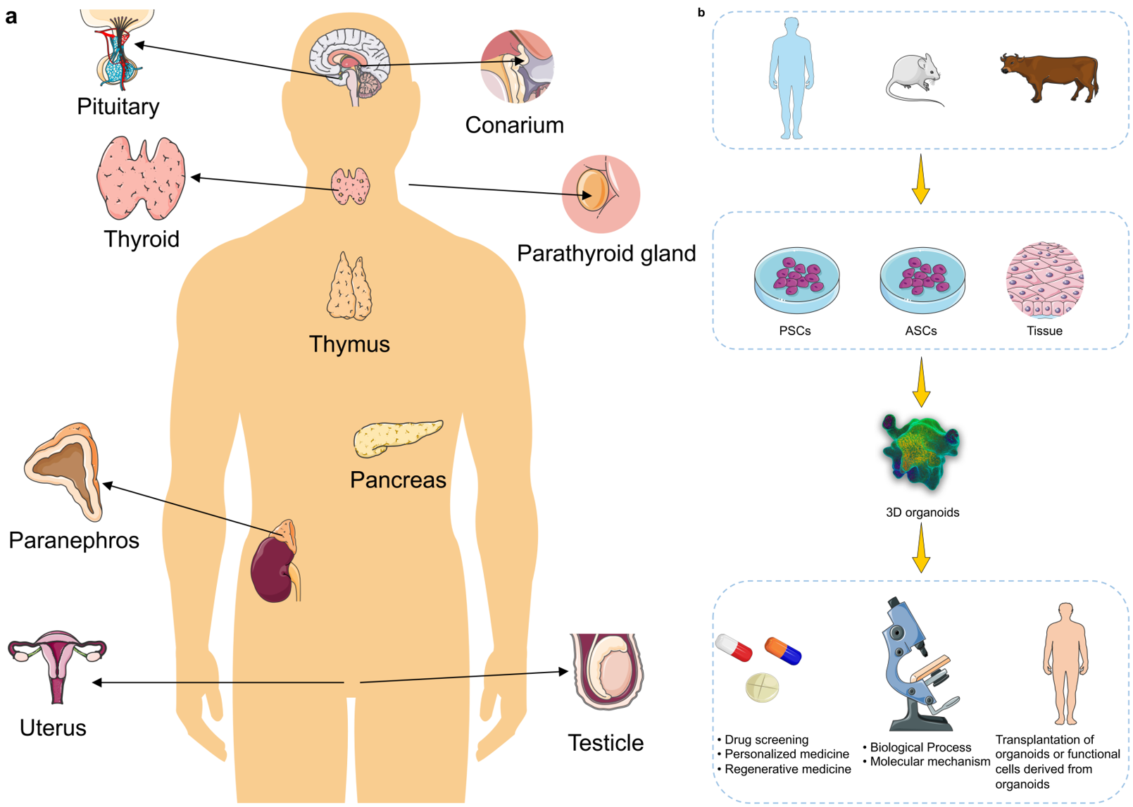
TY  - JOUR
AU  - Liu, Jiayu
AU  - Li, Peiting
PY  - 2026
DA  - 2026/02/06
TI  - The Use of Organoid Models of Endocrine Diseases: Research Progress and Potential
JO  - Oncology Communications
T2  - Oncology Communications
JF  - Oncology Communications
VL  - 1
IS  - 1
SP  - 8
EP  - 19
DO  - 10.62762/OC.2025.777438
UR  - https://www.icck.org/article/abs/OC.2025.777438
KW  - organoids
KW  - 3D culture
KW  - endocrine diseases
KW  - translational applications
KW  - disease modeling
KW  - tumor
AB  - The absence of robust and reliable $in \, vitro$ models that can accurately recapitulate the biological characteristics of many mammalian tissues and disease states represents a major barrier to both basic and translational research, owing to limited sample availability and ethical concerns. Stem cell-derived self-assembling three-dimensional (3D) organoids can replicate key structural and functional aspects of organs in a more physiologically relevant manner than traditional 2D models, thus providing a superior platform for simulating human physiology and pathology. To date, researchers have developed organoid models for a variety of endocrine tissues and their associated diseases (including pancreatic, pituitary, thyroid, adrenal tumors, etc.), offering invaluable tools for studying complex endocrine disorders. Such organoid models have significantly enhanced the accuracy and translational potential of research in disease modeling, drug screening, and regenerative medicine. Looking forward, the integration of bioengineering and multi-omics analyses with next-generation organoid models holds great promise for unraveling disease mechanisms and advancing precision medicine.
SN  - pending
PB  - Institute of Central Computation and Knowledge
LA  - English
ER  - 
BibTeX Format
Compatible with LaTeX, BibTeX, and other reference managers
BibTeX format data for LaTeX and reference managers
@article{Liu2026The,
  author = {Jiayu Liu and Peiting Li},
  title = {The Use of Organoid Models of Endocrine Diseases: Research Progress and Potential},
  journal = {Oncology Communications},
  year = {2026},
  volume = {1},
  number = {1},
  pages = {8-19},
  doi = {10.62762/OC.2025.777438},
  url = {https://www.icck.org/article/abs/OC.2025.777438},
  abstract = {The absence of robust and reliable \$in \, vitro\$ models that can accurately recapitulate the biological characteristics of many mammalian tissues and disease states represents a major barrier to both basic and translational research, owing to limited sample availability and ethical concerns. Stem cell-derived self-assembling three-dimensional (3D) organoids can replicate key structural and functional aspects of organs in a more physiologically relevant manner than traditional 2D models, thus providing a superior platform for simulating human physiology and pathology. To date, researchers have developed organoid models for a variety of endocrine tissues and their associated diseases (including pancreatic, pituitary, thyroid, adrenal tumors, etc.), offering invaluable tools for studying complex endocrine disorders. Such organoid models have significantly enhanced the accuracy and translational potential of research in disease modeling, drug screening, and regenerative medicine. Looking forward, the integration of bioengineering and multi-omics analyses with next-generation organoid models holds great promise for unraveling disease mechanisms and advancing precision medicine.},
  keywords = {organoids, 3D culture, endocrine diseases, translational applications, disease modeling, tumor},
  issn = {pending},
  publisher = {Institute of Central Computation and Knowledge}
}

Article Metrics
Citations:

Crossref

0

Scopus

0

Web of Science

0
Article Access Statistics:
Views: 2
PDF Downloads: 0

Publisher's Note
ICCK stays neutral with regard to jurisdictional claims in published maps and institutional affiliations.

Rights and Permissions
CC BY Copyright © 2026 by the Author(s). Published by Institute of Central Computation and Knowledge. This article is an open access article distributed under the terms and conditions of the Creative Commons Attribution (CC BY) license (https://creativecommons.org/licenses/by/4.0/), which permits use, sharing, adaptation, distribution and reproduction in any medium or format, as long as you give appropriate credit to the original author(s) and the source, provide a link to the Creative Commons licence, and indicate if changes were made.
Oncology Communications

Oncology Communications

ISSN: pending (Online)

Email: [email protected]

Portico

Portico

All published articles are preserved here permanently:
https://www.portico.org/publishers/icck/