

Robust Skin Lesion Segmentation Using a Hybrid CNN Model and Compound Scaling



Article Information
Abstract
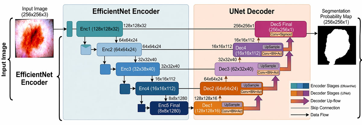
Medical image processing by computers has become an area of growing importance, with researchers shifting their attention to various groundbreaking state-of-the-art models that aim to ease the pressure on doctors. Notwithstanding, image segmentation and feature extraction are critical yet challenging tasks in clinical diagnostics, hindered by issues such as noise and artifacts, limited annotated data, and the need for high computational efficiency. This research focuses on developing an EfficientNet-U-Net model, which aims to improve medical image segmentation for detecting skin lesions utilizing the International Skin Imaging Collaboration Challenge (ISIC) 2018 dataset. The model combines the EfficientNet encoder, optimized for proficient feature extraction via compound scaling, with a U-Net-style decoder that uses transposed convolutions and skip connections to preserve spatial information. The approach also utilizes a hybrid loss function that combines Dice and Binary Cross-Entropy, as well as an adaptive learning rate schedule. Experimental results indicate that the proposed model performs well for the fully automated identification and delineation of skin lesions with the potential for clinical use. The proposed model achieves performance comparable to that of existing skin lesion segmentation methods.
Graphical Abstract
Keywords
Data Availability Statement
Funding
Conflicts of Interest
AI Use Statement
Ethical Approval and Consent to Participate
References
- Arnold, M., Singh, D., Laversanne, M., Vignat, J., Vaccarella, S., Meheus, F., ... & Bray, F. (2022). Global burden of cutaneous melanoma in 2020 and projections to 2040. JAMA dermatology, 158(5), 495-503.
[CrossRef] [Google Scholar] - Liu, M., Wu, M., Liu, X., Zhou, J., Lan, Y., Zhang, H., ... & Li, J. (2023). Assessing the quality of care for skin malignant melanoma on a global, regional, and national scale: a systematic analysis of the global burden of disease study from 1990 to 2019. Archives of Dermatological Research, 315(10), 2893-2904.
[CrossRef] [Google Scholar] - Mirikharaji, Z., Abhishek, K., Bissoto, A., Barata, C., Avila, S., Valle, E., ... & Hamarneh, G. (2023). A survey on deep learning for skin lesion segmentation. Medical Image Analysis, 88, 102863.
[CrossRef] [Google Scholar] - Mustafa, S., Jaffar, A., Rashid, M., Akram, S., & Bhatti, S. M. (2025). Deep learning-based skin lesion analysis using hybrid ResUNet++ and modified AlexNet-Random Forest for enhanced segmentation and classification. PloS one, 20(1), e0315120.
[CrossRef] [Google Scholar] - Manzoor, K., Gilal, N. U., Agus, M., & Schneider, J. (2025). Dual-stage segmentation and classification framework for skin lesion analysis using deep neural network. Digital health, 11, 20552076251351858.
[CrossRef] [Google Scholar] - Schmidt, A., Morales-Alvarez, P., & Molina, R. (2023, October). Probabilistic modeling of inter-and intra-observer variability in medical image segmentation. In 2023 IEEE/CVF International Conference on Computer Vision (ICCV) (pp. 21040-21049). IEEE.
[CrossRef] [Google Scholar] - Abdelrahman, A., & Viriri, S. (2023). EfficientNet family U-Net models for deep learning semantic segmentation of kidney tumors on CT images. Frontiers in Computer Science, 5, 1235622.
[CrossRef] [Google Scholar] - Bachanek, S., Wuerzberg, P., Biggemann, L., Janssen, T. Y., Nietert, M., Lotz, J., ... & Uhlig, J. (2025). Renal tumor segmentation, visualization, and segmentation confidence using ensembles of neural networks in patients undergoing surgical resection. European Radiology, 35(4), 2147-2156.
[CrossRef] [Google Scholar] - Manole, I., & Tiplica, G. S. (2024). Integrating Artificial Intelligence in dermatology: progress, challenges and perspectives. Romanian Medical Journal, 71(2), 183.
[CrossRef] [Google Scholar] - Litjens, G., Kooi, T., Bejnordi, B. E., Setio, A. A. A., Ciompi, F., Ghafoorian, M., ... & Sánchez, C. I. (2017). A survey on deep learning in medical image analysis. Medical image analysis, 42, 60-88.
[CrossRef] [Google Scholar] - Minaee, S., Boykov, Y., Porikli, F., Plaza, A., Kehtarnavaz, N., & Terzopoulos, D. (2021). Image segmentation using deep learning: A survey. IEEE transactions on pattern analysis and machine intelligence, 44(7), 3523-3542.
[CrossRef] [Google Scholar] - Ronneberger, O., Fischer, P., & Brox, T. (2015, October). U-net: Convolutional networks for biomedical image segmentation. In International Conference on Medical image computing and computer-assisted intervention (pp. 234-241). Cham: Springer international publishing.
[CrossRef] [Google Scholar] - Esteva, A., Kuprel, B., Novoa, R. A., Ko, J., Swetter, S. M., Blau, H. M., & Thrun, S. (2017). Dermatologist-level classification of skin cancer with deep neural networks. Nature, 542(7639), 115-118.
[CrossRef] [Google Scholar] - Isensee, F., Jaeger, P. F., Kohl, S. A., Petersen, J., & Maier-Hein, K. H. (2021). nnU-Net: a self-configuring method for deep learning-based biomedical image segmentation. Nature methods, 18(2), 203-211.
[CrossRef] [Google Scholar] - Su, R., Zhang, D., Liu, J., & Cheng, C. (2021). Msu-net: Multi-scale u-net for 2d medical image segmentation. Frontiers in Genetics, 12, 639930.
[CrossRef] [Google Scholar] - Tan, M., & Le, Q. (2019). EfficientNet: Rethinking model scaling for convolutional neural networks. International Conference on Machine Learning, 6105-6114. https://proceedings.mlr.press/v97/tan19a.html?ref=ji
[Google Scholar] - Jha, D., Riegler, M. A., Johansen, D., Halvorsen, P., & Johansen, H. D. (2020, July). Doubleu-net: A deep convolutional neural network for medical image segmentation. In 2020 IEEE 33rd International symposium on computer-based medical systems (CBMS) (pp. 558-564). IEEE.
[CrossRef] [Google Scholar] - Trinh, M. N., Nguyen, V. D., Pham, V. T., & Tran, T. T. (2023). An EffcientNet-encoder U-Net joint residual refinement module with Tversky–Kahneman Baroni–Urbani–Buser loss for biomedical image segmentation. Biomedical Signal Processing and Control, 83, 104631.
[CrossRef] [Google Scholar] - Zhou, Z., Siddiquee, M. M. R., Tajbakhsh, N., & Liang, J. (2019). Unet++: Redesigning skip connections to exploit multiscale features in image segmentation. IEEE transactions on medical imaging, 39(6), 1856-1867.
[CrossRef] [Google Scholar] - Ummadi, V. (2022). U-Net and its variants for Medical Image Segmentation: A short review. arXiv preprint arXiv:2204.08470.
[CrossRef] [Google Scholar] - Abedalla, A., Abdullah, M., Al-Ayyoub, M., & Benkhelifa, E. (2021). Chest X-ray pneumothorax segmentation using U-Net with EfficientNet and ResNet architectures. PeerJ Computer Science, 7, e607.
[CrossRef] [Google Scholar] - Vong, K. M., & Dinh, T. B. (2021). Pneumothorax segmentation in chest X-rays using UNet++ and EfficientNet. 2021 IEEE EMBS International Conference on Biomedical and Health Informatics (BHI), 1-4.
[CrossRef] [Google Scholar] - Ruan, J., Xie, M., Gao, J., Liu, T., & Fu, Y. (2023, October). Ege-unet: an efficient group enhanced unet for skin lesion segmentation. In International conference on medical image computing and computer-assisted intervention (pp. 481-490). Cham: Springer Nature Switzerland.
[CrossRef] [Google Scholar] - Lesion Segmentation on ISIC 2018 (Ranking from SOTA). (n.d.). Retrieved March 2025, from Papers with Code. https://paperswithcode.com/sota/lesion-segmentation-on-isic-2018
[Google Scholar] - Wang, Q., Wu, B., Zhu, P., Li, P., Zuo, W., & Hu, Q. (2020, June). ECA-Net: Efficient channel attention for deep convolutional neural networks. In 2020 IEEE/CVF conference on computer vision and pattern recognition (CVPR) (pp. 11531-11539). IEEE.
[CrossRef] [Google Scholar] - Li, X., Zhu, W., Dong, X., Dumitrascu, O. M., & Wang, Y. (2025, April). EViT-Unet: U-Net like efficient vision transformer for medical image segmentation on mobile and edge devices. In 2025 IEEE 22nd International Symposium on Biomedical Imaging (ISBI) (pp. 1-5). IEEE.
[CrossRef] [Google Scholar] - Chen, J., Lu, Y., Yu, Q., Luo, X., Adeli, E., Wang, Y., & Zhou, Y. (2021). TransUNet: Transformers make strong encoders for medical image segmentation. arXiv preprint arXiv:2102.04306.
[CrossRef] [Google Scholar] - Du, G., Cao, X., Liang, J., Chen, X., & Zhan, Y. (2020). Medical image segmentation based on U-net: A review. Journal of Imaging Science & Technology, 64(2), 1.
[CrossRef] [Google Scholar] - Siddique, N., Sidike, P., Elkin, C., & Devabhaktuni, V. (2020). U-Net and its variants for medical image segmentation: theory and applications. arXiv preprint arXiv:2011.01118.
[CrossRef] [Google Scholar] - Park, M., Oh, S., Park, J., Jeong, T., & Yu, S. (2025). ES-UNet: efficient 3D medical image segmentation with enhanced skip connections in 3D UNet. BMC medical imaging, 25(1), 327.
[CrossRef] [Google Scholar] - Lee, H., Park, J., & Hwang, J. Y. (2020). Channel attention module with multiscale grid average pooling for breast cancer segmentation in an ultrasound image. IEEE transactions on ultrasonics, ferroelectrics, and frequency control, 67(7), 1344-1353.
[CrossRef] [Google Scholar] - Zhang, C., Deng, X., & Ling, S. H. (2024). Next-gen medical imaging: U-Net evolution and the rise of transformers. Sensors, 24(14), 4668.
[CrossRef] [Google Scholar] - Asadi-Aghbolaghi, M., Azad, R., Fathy, M., & Escalera, S. (2020). Multi-level context gating of embedded collective knowledge for medical image segmentation. arXiv preprint arXiv:2003.05056.
[CrossRef] [Google Scholar] - Tiwary, P. K., Johri, P., Katiyar, A., & Chhipa, M. K. (2025). Deep learning-based MRI brain tumor segmentation with EfficientNet-enhanced UNet. IEEE Access, 13, 54920-54937.
[CrossRef] [Google Scholar] - Rais, K., Amroune, M., Haouam, M. Y., & Benmachiche, A. (2025). Enhancing medical image analysis through geometric and photometric transformations. arXiv preprint arXiv:2501.13643.
[CrossRef] [Google Scholar] - Raghu, M., Zhang, C., Kleinberg, J., & Bengio, S. (2019). Transfusion: Understanding transfer learning for medical imaging. Advances in neural information processing systems, 32.
[Google Scholar] - Tschandl, P., Rosendahl, C., & Kittler, H. (2018). The HAM10000 dataset, a large collection of multi-source dermatoscopic images of common pigmented skin lesions. Scientific Data, 5, 180161.
[CrossRef] [Google Scholar] - Chowdary, G. J., Yathisha, G. V. S. N. D., Suganya, G., & Premalatha, M. (2021). Automated skin lesion segmentation using multi-scale feature extraction scheme and dual-attention mechanism. arXiv preprint arXiv:2111.08708.
[CrossRef] [Google Scholar] - Benčević, M., Galić, I., Habijan, M., & Babin, D. (2021). Training on polar image transformations improves biomedical image segmentation. IEEE access, 9, 133365-133375.
[CrossRef] [Google Scholar] - Tang, F., Xu, Z., Huang, Q., Wang, J., Hou, X., Su, J., & Liu, J. (2023, October). DuAT: Dual-aggregation transformer network for medical image segmentation. In Chinese conference on pattern recognition and computer vision (PRCV) (pp. 343-356). Singapore: Springer Nature Singapore.
[CrossRef] [Google Scholar] - Wang, J., Wei, L., Wang, L., Zhou, Q., Zhu, L., & Qin, J. (2021, September). Boundary-aware transformers for skin lesion segmentation. In International conference on medical image computing and computer-assisted intervention (pp. 206-216). Cham: Springer International Publishing.
[CrossRef] [Google Scholar]
Cite This Article
TY  - JOUR
AU  - Asante-Mensah, Maame G.
AU  - Haruna, Charles Roland
AU  - Atsu, Stephen Julius
AU  - Amofa, Sandro Kwame
AU  - Acquah, Akua Gyanba Morgan
PY  - 2026
DA  - 2026/10/20
TI  - Robust Skin Lesion Segmentation Using a Hybrid CNN Model and Compound Scaling
JO  - ICCK Transactions on Machine Intelligence
T2  - ICCK Transactions on Machine Intelligence
JF  - ICCK Transactions on Machine Intelligence
VL  - 2
IS  - 4
SP  - 213
EP  - 223
DO  - 10.62762/TMI.2026.565544
UR  - https://www.icck.org/article/abs/TMI.2026.565544
KW  - medical imaging
KW  - EfficientNet-UNet
KW  - skip connection
KW  - skin lesion
KW  - image segmentation
AB  - Medical image processing by computers has become an area of growing importance, with researchers shifting their attention to various groundbreaking state-of-the-art models that aim to ease the pressure on doctors. Notwithstanding, image segmentation and feature extraction are critical yet challenging tasks in clinical diagnostics, hindered by issues such as noise and artifacts, limited annotated data, and the need for high computational efficiency. This research focuses on developing an EfficientNet-U-Net model, which aims to improve medical image segmentation for detecting skin lesions utilizing the International Skin Imaging Collaboration Challenge (ISIC) 2018 dataset. The model combines the EfficientNet encoder, optimized for proficient feature extraction via compound scaling, with a U-Net-style decoder that uses transposed convolutions and skip connections to preserve spatial information. The approach also utilizes a hybrid loss function that combines Dice and Binary Cross-Entropy, as well as an adaptive learning rate schedule. Experimental results indicate that the proposed model performs well for the fully automated identification and delineation of skin lesions with the potential for clinical use. The proposed model achieves performance comparable to that of existing skin lesion segmentation methods.
SN  - 3068-7403
PB  - Institute of Central Computation and Knowledge
LA  - English
ER  -
@article{AsanteMensah2026Robust,
  author = {Maame G. Asante-Mensah and Charles Roland Haruna and Stephen Julius Atsu and Sandro Kwame Amofa and Akua Gyanba Morgan Acquah},
  title = {Robust Skin Lesion Segmentation Using a Hybrid CNN Model and Compound Scaling},
  journal = {ICCK Transactions on Machine Intelligence},
  year = {2026},
  volume = {2},
  number = {4},
  pages = {213-223},
  doi = {10.62762/TMI.2026.565544},
  url = {https://www.icck.org/article/abs/TMI.2026.565544},
  abstract = {Medical image processing by computers has become an area of growing importance, with researchers shifting their attention to various groundbreaking state-of-the-art models that aim to ease the pressure on doctors. Notwithstanding, image segmentation and feature extraction are critical yet challenging tasks in clinical diagnostics, hindered by issues such as noise and artifacts, limited annotated data, and the need for high computational efficiency. This research focuses on developing an EfficientNet-U-Net model, which aims to improve medical image segmentation for detecting skin lesions utilizing the International Skin Imaging Collaboration Challenge (ISIC) 2018 dataset. The model combines the EfficientNet encoder, optimized for proficient feature extraction via compound scaling, with a U-Net-style decoder that uses transposed convolutions and skip connections to preserve spatial information. The approach also utilizes a hybrid loss function that combines Dice and Binary Cross-Entropy, as well as an adaptive learning rate schedule. Experimental results indicate that the proposed model performs well for the fully automated identification and delineation of skin lesions with the potential for clinical use. The proposed model achieves performance comparable to that of existing skin lesion segmentation methods.},
  keywords = {medical imaging, EfficientNet-UNet, skip connection, skin lesion, image segmentation},
  issn = {3068-7403},
  publisher = {Institute of Central Computation and Knowledge}
}
Article Metrics
Publisher's Note
ICCK stays neutral with regard to jurisdictional claims in published maps and institutional affiliations.
Rights and Permissions


Portico
